
 |
小肠袢间的粘连,腹部手术后的常见并发症。腹膜炎也可能发生更广泛的粘连。 This is an adhesion between loops of small intestine. Such adhesions are typical following abdominal surgery. More diffuse adhesions may also form following peritonitis. |
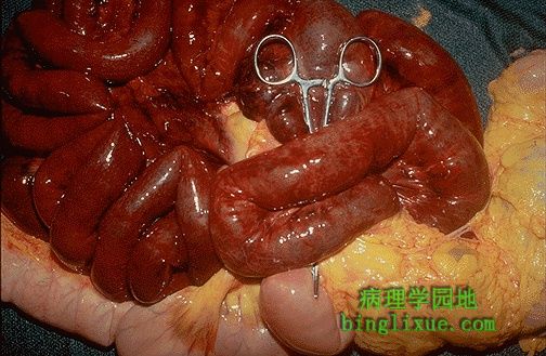 |
与明亮的、粉红色的、有活力的肠相比梗死的小肠呈暗红色,梗死是由于手术后发生粘连导致的绞榨性疝。梗死的肠已经失去了血液供应。 The dark red infarcted small intestine contrasts with the light pink viable bowel. The forceps extend through an internal hernia in which a loop of bowel and mesentery has been caught. This is one complication of adhesions from previous surgery. The trapped bowel has lost its blood supply. |
 |
盲肠扭转,肠扭转是肠的扭曲缠绕。成年人肠扭转较常见,发生在小肠(围绕着扭曲的肠系膜)和结肠(发生在乙状结肠或是盲肠,它不固定)机会是均等的。儿童肠扭转常发生于小肠。 This is an example of cecal volvulus. Volvulus is a twisting of the bowel. Volvulus is most common in adults, where it occurs with equal frequency in small intestine (around a twisted mesentery) and colon (in either sigmoid or cecum which are more mobile). In very young children, volvulus almost always happens in the small intestine. |
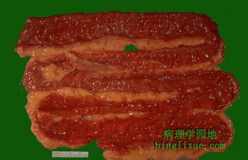 |
小肠黏膜显示明显充血 ,是局部缺血性肠炎的结果。局部缺血常见于由心衰、大量失血、血管受阻(见于肠疝、肠扭转或肠套叠)所导致的低血压(休克)。如果血液供应没有迅速恢复,肠就会梗死。 The small intestinal mucosa demonstrates marked hyperemia as a result of ischemic enteritis. Such ischemia most often results from hypotension (shock) from cardiac failure, from marked blood loss, or from loss of blood supply from mechanical obstruction (as with the bowel incarcerated in a hernia or with volvulus or intussusception). If the blood supply is not quickly restored, the bowel will infarct. |
 |
早期的局部缺血性肠炎肠绒毛近距离检查。通常,肠很难由于动脉粥样硬化血管狭窄或血栓栓塞而发生梗死,因为有广泛吻合支存在。大多数肠局部缺血和梗死的病例,是由于血压过低和心输出量降低引起的。 On closer inspection, early ischemic enteritis involves the tips of the villi. In general, bowel is hard to infarct from atherosclerotic vascular narrowing or thromboembolization because of the widely anastomosing blood supply. Thus, most cases of bowel ischemia and infarction result from generalized hypotension and decreased cardiac output. |
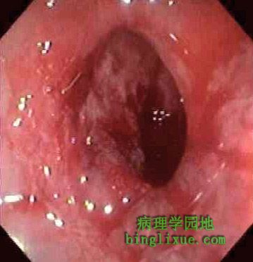 |
有少量分泌物覆盖的缺血性结肠炎的结肠镜检图在下面可见。 A colonoscopic view of ischemic colitis with minimal overlying exudate is shown. |
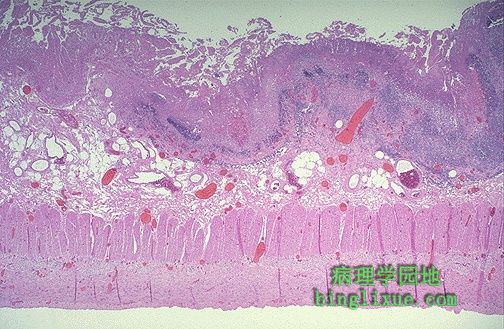 |
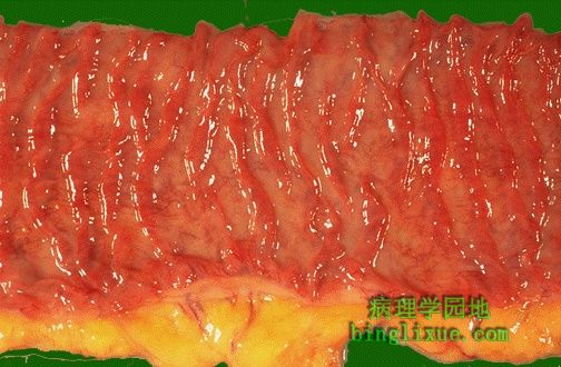
肠黏膜表面显示有早期坏死,从黏膜向黏膜下层和血管壁肌层都有充血。然而,黏膜下层和肌层仍然保持完整。 The mucosal surface of the bowel seen here shows early necrosis with hyperemia extending all the way from mucosa to submucosal and muscular wall vessels. The submucosa and muscularis, however, are still intact. |
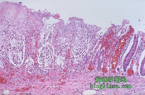 |
较高倍镜下,局部缺血性急性肠炎显示更早期坏死,小肠黏膜有出血。 At higher magnification with more advanced necrosis, the small intestinal mucosa shows hemorrhage with acute inflammation in this case of ischemic enteritis. |
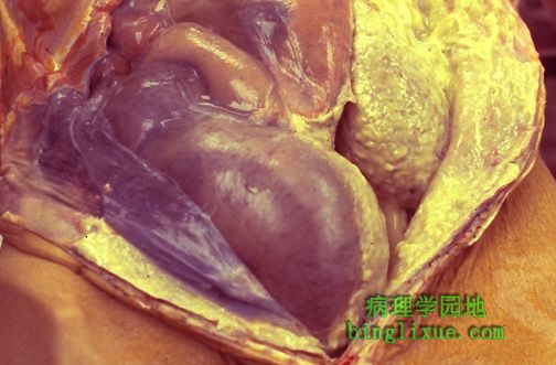 |
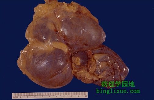
胃肠道穿孔(食管下段到结肠)能导致腹膜炎,如图尸检所见。浓密的黄*色脓性渗出物覆盖在腹膜表面。卵巢癌引起乙状结肠的梗阻、穿孔。骨盆中可见明显扩张和灰黑色的乙状结肠。 Perforation of GI tract (from lower esophagus to colon) can result in a peritonitis as seen here at autopsy. A thick yellow purulent exudate covers peritoneal surfaces. An ovarian carcinoma caused sigmoid colonic obstruction (the sigmoid is the markedly dilated grey-black bowel in the pelvis seen here) with perforation. |
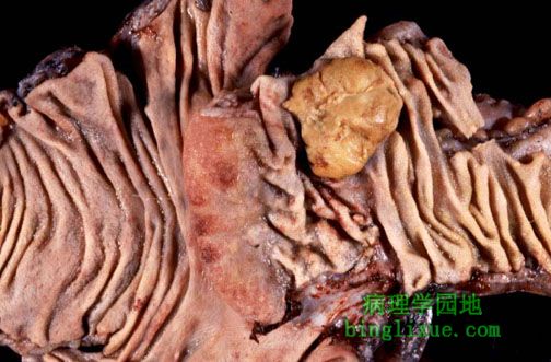 |
小肠肿瘤不常见。良性肿瘤包括平滑肌瘤、纤维瘤、神经纤维瘤和脂肪瘤。图示回盲瓣部见浅黄*色的肿块,属类癌。虽然,良性肿瘤很少大到阻塞肠腔,但很多有黏膜下层破坏。 Neoplasms of the small intestine are uncommon. Benign tumors can include leiomyomas, fibromas, neurofibromas, and lipomas. Seen here at the ileocecal valve is another tumor that has a faint yellowish color. This is a carcinoid tumor. Most benign tumors are incidental submucosal lesions, though rarely they can be large enough to obstruct the lumen. |