
 |
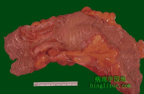
左侧显示结肠坚硬腺癌的浸润性包块,是发生在降结肠的典型的腺癌。包块影响的结果是大便习惯的改变。 The encircling mass of firm adenocarcinoma in this colon at the left is typical for adenocarcinomas arising in the descending colon. A change in stool or bowel habits can be created by the mass effect. |
 |
结肠镜检,可见一个真菌样生长的溃疡面。 By colonoscopy, a fungating, ulcerating mass is seen in the views below. |
 |
结肠浸润型腺癌CT图像。 This CT image of the abdomen demonstrates an encircling mass involving the colon. This is a colonic adenocarcinoma. |
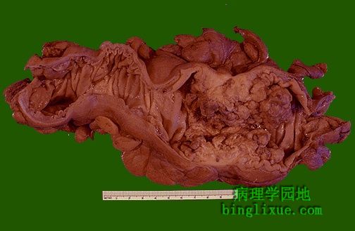 |
结肠腺癌,呈外生性生长,因此梗阻就成了它的一个并发症(通常是部分性梗阻)。 Here is another example of an adenocarcinoma of colon. This cancer is more exophytic in its growth pattern. Thus, one of the complications of a carcinoma is obstruction (usually partial). |
 |
直肠腺癌的结肠镜检图显示溃疡型肿块。 Colonoscopic views of another ulcerating mass, a rectal adenocarcinoma, are seen below. |
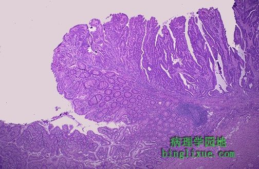 |
图示绒毛状腺瘤恶的癌的边缘。肿瘤腺体长并呈叶状,与绒毛状腺瘤所见相似。生长最初是外生性的(朝向腔内),不见浸润。肿瘤的分级和分期由外科病理学家检查肿瘤的多个组织部位而定。 The edge of the carcinoma arising in the villous adenoma is seen here. The neoplastic glands are long and frond-like, similar to those seen in a villous adenoma. The growth is primarily exophytic (outward into the lumen) and invasion is not seen at this point. Grading and staging of the tumor is done by the surgical pathologist who will examine multiple histologic sections of the tumor. |
 |
显微镜下,中分化结肠腺癌。有腺样结构,但腺体不规则、密集分布。很多腺体有含有兰色黏液的腺腔。 Microscopically, a moderately differentiated adenocarcinoma of colon is seen here. There is still a glandular configuration, but the glands are irregular and very crowded. Many of them have lumens containing bluish mucin. |
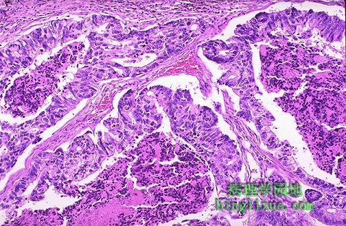 |
腺癌,腺体更大,而且充满坏死的碎片。 Here is an adenocarcinoma in which the glands are much larger and filled with necrotic debris. |
 |
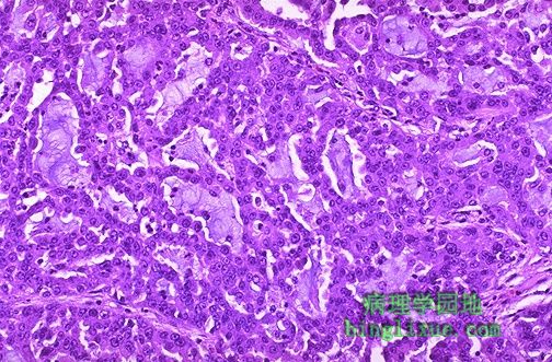
高倍镜下腺癌腺体见大量密集深染异型性明显的细胞核。不见正常的杯状细胞。 At high magnification, the neoplastic glands of adenocarcinoma have crowded nuclei with hyperchromatism and pleomorphism. No normal goblet cells are seen. |
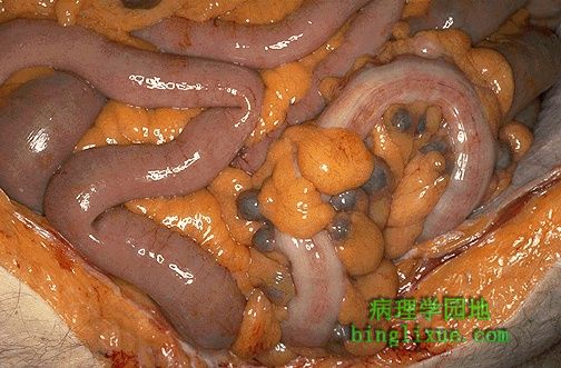 |
右边的乙状结肠在颜色上比临近的小肠浅,有结肠带。乙状结肠的突出物是多个圆形的蓝灰色的憩室。憩室发生在结肠比小肠更常见。易于发生在左半结肠。它们常发生在发达国家人群中,他们的食物中纤维素含量低。 The sigmoid colon at the right appears lighter in color than the adjacent small intestine and has a band of taenia coli muscle running longitudinally. Protruding from the sigmoid colon are multiple rounded bluish-gray diverticula. Diverticula are much more common in the colon than in small intestine, and they are more common in the left colon, and they are more common in persons living in developed nations in which the usual diet has less fiber. |