
 |
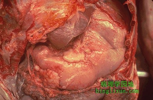
尸检胸腹腔被剖开,右侧可见胃和肝下缘。胃腺癌已浸润胃壁,表面呈现不规则的褐色肿块,胃腺癌广泛浸润引起幽门梗阻。 At autopsy, the thoracic cavity and abdominal cavity are both opened to reveal the stomach just to the right and below the edge of liver in this photograph. Gastric adenocarcinoma has infiltrated through the wall and appears on the surface as irregular tan masses. The extensive tumor in this case caused gastric outlet obstruction. |
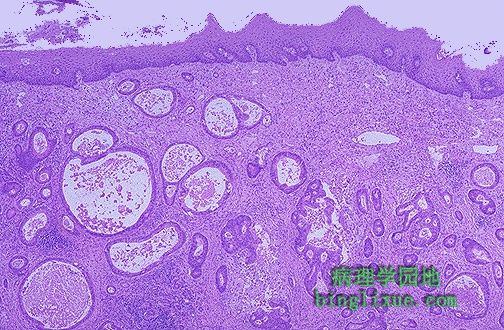 |
中分化胃腺癌向上浸润,浸润到食管的鳞状上皮黏膜下层,肿瘤腺体大小不一。 A moderately differentiated gastric adenocarcinoma is infiltrating up and into the submucosa below the squamous mucosa of the esophagus. The neoplastic glands are variably sized. |
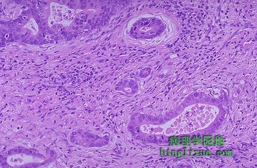 |
高倍镜下,胃腺癌腺体细胞分裂象多见,核质比增加,着色过深。浸润腺体后结缔组织增生。 At higher magnification, the neoplastic glands of gastric adenocarcinoma demonstrate mitoses, increased nuclear/cytoplasmic ratios, and hyperchromatism. There is a desmoplastic stromal reaction to the infiltrating glands. |
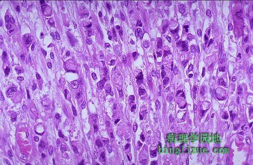 |
高倍镜下,胃腺癌分化较差,腺体已不可见。相反,可见异型性明显的的肿瘤细胞的成行排列。很多肿瘤细胞有明显的黏液空泡。 At high power, this gastric adenocarcinoma is so poorly differentiated that glands are not visible. Instead, rows of infiltrating neoplastic cells with marked pleomorphism are seen. Many of the neoplastic cells have clear vacuoles of mucin. |
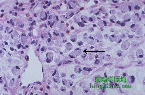 |
如图腺癌表现为印戒细胞癌,细胞里充满黏液空泡把核挤向一边,如箭头所示。 This is a signet ring cell pattern of adenocarcinoma in which the cells are filled with mucin vacuoles that push the nucleus to one side, as shown at the arrow. |
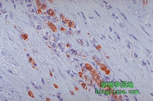 |
抗体对细胞角蛋白免疫过氧化物酶染色,低分化肿瘤细胞呈阳性,如图所示肿瘤细胞已浸润胃壁,细胞角蛋白染色主要用于源于上皮的恶性肿瘤(癌)。 This is an immunoperoxidase stain with antibody to cytokeratin, which is positive in the poorly differentiated neoplastic cells seen here infiltrating through the gastric wall. Cytokeratin staining is typical for neoplasms of epithelial origin (carcinomas). |
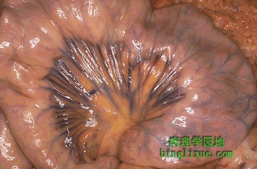 |
可见由肠系膜固着的肠袢。注意肠静脉分布,动脉经由同一区域。这样,有一广泛的动脉网向肠供血,使其很难梗死。同样,广泛的静脉管道汇合成门静脉入肝。 Seen here is a loop of bowel attached via the mesentery. Note the extent of the veins. Arteries run in the same location. Thus, there is an extensive anastomosing arterial blood supply to the bowel, making it more difficult to infarct. Also, the extensive venous drainage is incorporated into the portal venous system heading to the liver. |
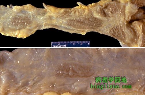 |
正常回肠末段外观,上图显示回盲瓣和黏膜上的几个较暗的椭圆形肠道集合淋巴结。下图显示肠道集合淋巴结(黏膜下层淋巴组织集中的地方)。皱褶不如在空肠那样明显。如下面的结肠镜检图清楚显示的一样。 This is the normal appearance of terminal ileum. In the upper frame, note the ileocecal valve, and several darker oval Peyer's patches are present on the mucosa. In the lower frame, a Peyer's patch, which is a concentration of submucosal lymphoid tissue, is present. Note the folds are not as prominent here as in the jejunum, as evidenced by the colonoscopic view below. |
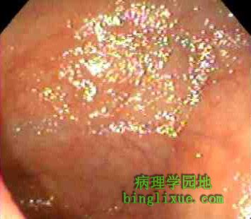 |
结肠镜检皱褶不明显。 |
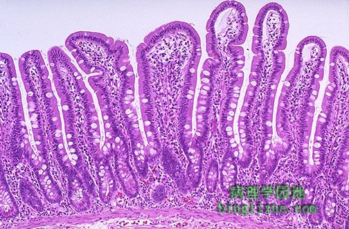 |
正常小肠黏膜,有较长绒毛,偶尔可见杯状细胞。绒毛为消化和吸收提供了很大的面积。 This is the normal appearance of small intestinal mucosa with long villi that have occasional goblet cells. The villi provide a large area for digestion and absorption. |