
 |
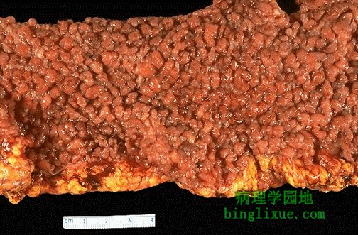
息肉病,结肠黏膜覆盖有很多小息肉。在这个特殊的病例中,有颅骨骨瘤,壶腹周围的腺癌和表皮囊肿。称为加德纳综合征(Gardner 综合征)。家族性腺瘤息肉病的遗传方式是常染色体显性遗传。 Here is another example of polyposis with numerous small polyps covering the colonic mucosa. In this particular case, there were osteomas of the skull, a periampullary adenocarcinoma, and epidermal inclusion cysts. Thus, this is a case of Gardner's syndrome. As with familial adenomatous polyposis, the inheritance pattern is autosomal dominant. |
 |
左侧为正常结肠黏膜,右侧为腺瘤性息肉(管状腺瘤)。腺体不规则,并有深染的、密集的细胞核 。肿块是良性的,分化很好的,与正常结肠组织相似。 A microscopic comparison of normal colonic mucosa on the left and that of an adenomatous polyp (tubular adenoma) on the right is seen here. The neoplastic glands are more irregular with darker (hyperchromatic) and more crowded nuclei. This neoplasm is benign and well-differentiated, as it still closely resembles the normal colonic structure. |
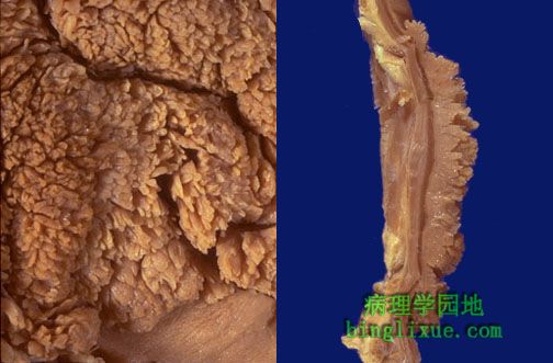 |
绒毛状腺瘤,左边显示表面,右边显示横切面。无蒂,比管状腺瘤(腺瘤息肉)要大。绒毛状腺瘤直径一般几厘米,可以达到10个厘米。 The gross appearance of a villous adenoma is shown above the surface at the left, and in cross section at the right. Note that this type of adenoma is sessile, rather than pedunculated, and larger than a tubular adenoma (adenomatous polyp). A villous adenoma averages several centimeters in diameter, and may be up to 10 cm. |
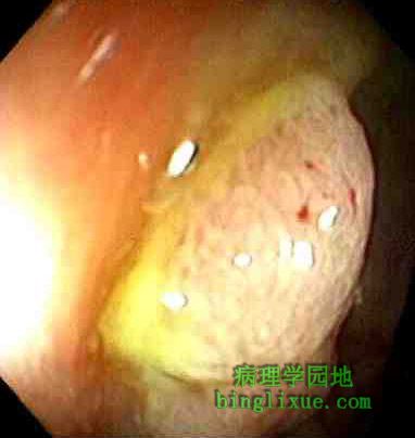 |
结肠镜检查,在下边可见一个无蒂的息肉。 On colonoscopy, a sessile polyp is seen below. |
 |
显微镜下,左侧见绒毛状腺瘤边缘,右侧见突破基底膜。菜花样外观是由于伸长的腺体结构被发育异常的上皮覆盖。虽然绒毛状腺瘤不如腺瘤息肉那么常见,但更有可能恶变为癌(大约40%的绒毛腺瘤)。 Microscopically, a villous adenoma is shown at its edge on the left, and projecting above the basement membrane at the right. The cauliflower-like appearance is due to the elongated glandular structures covered by dysplastic epithelium. Though villous adenomas are less common than adenomatous polyps, they are much more likely to have invasive carcinoma in them (about 40% of villous adenomas). |
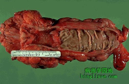 |
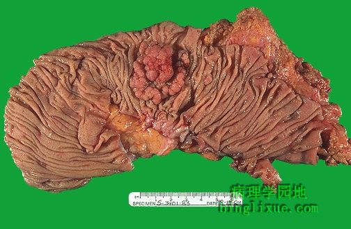
直肠上部和乙状结肠下部浸润型腺癌。溃疡边缘有隆起的肿瘤块。会引起出血,可以通过粪便检查出来。右侧显示正常黏膜。肿瘤环绕结肠四周浸润肠壁。分期是根据浸润和破坏结肠的程度。 An encircling adenocarcinoma of the rectosigmoid region is seen here. There is a heaped up margin of tumor at each side with a central area of ulceration. This produces the bleeding that allows detection through a stool guaiac test. Normal mucosa appears at the right. The tumor encircles the colon and infiltrates into the wall. Staging is based upon the degree of invasion into and through the wall. |
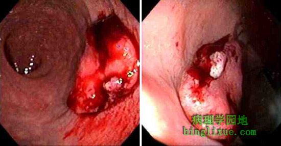 |
小的直肠腺癌的结肠镜检图,表面有一溃烂。 The colonoscopic views of a smaller rectal adenocarcinoma, but still with an ulcerated surface, are shown here. |
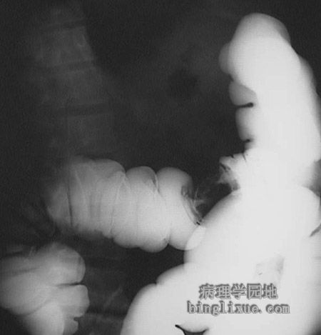 |
The barium enema technique instills the radiopaque barium sulfate into the colon, producing a contrast with the wall of the colon that highlights any masses present. In this case, the classic "apple core" lesion is present, representing an encircling adenocarcinoma that constricts the lumen. 钡剂灌肠显示结肠有一明显肿块,与高亮区形成鲜明对比。此病例表现为苹果核样病变,是由于浸润型结肠癌浸润肠壁四周,使管腔狭窄甚至闭塞。 |
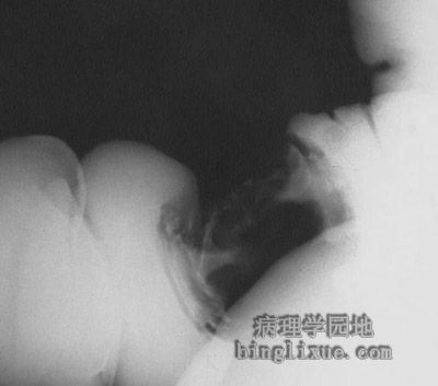 |
浸润型结肠癌钡剂灌肠放大图。 Below is a higher magnification of the radiograph above. |
 |
绒毛状腺瘤恶变的腺癌,肿瘤呈息肉状微红色,肿瘤表现出血使得粪便潜血检查呈阳性,该肿瘤块位于乙状结肠,指诊触不到,但结肠镜检病变清楚可见。 This is an adenocarcinoma arising in a villous adenoma. The surface of the neoplasm is polypoid and reddish pink. Hemorrhage from the surface of the tumor creates a guaiac positive stool. This neoplasm was located in the sigmoid colon, just out of reach of digital examination, but easily visualized with sigmoidoscopy. |