
 |
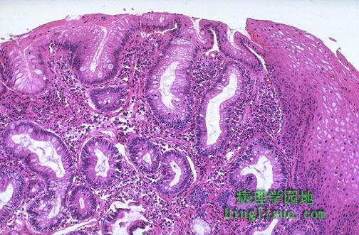
Barrett食管,多见于慢性返流性食管炎。胃食管交界处上方有胃黏膜上皮(柱状上皮)。左为柱状上皮,右为鳞状上皮。图示典型的Barrett食管黏膜,因有肠上皮化生(柱状上皮内也可见杯状细胞。) Another cause for inflammation is a so-called "Barrett's esophagus" in which there is gastric-type mucosa above the gastroesophageal junction. Note the columnar epithelium to the left and the squamous epithelium at the right. This is "typical" Barrett's mucosa, because there is intestinal metaplasia as well (note the goblet cells in the columnar mucosa). |
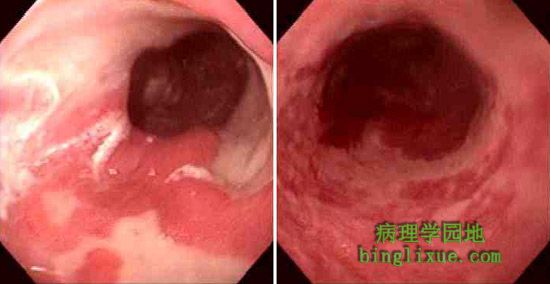 |
胃镜显示Barrett食管食管下段的黏膜红斑,也可见孤立的正常灰白色的食管鳞状上皮。 These two endoscopic views demonstrate Barrett esophagus areas of mucosal erythema of the lower esophagus, with islands of normal pale esophageal squamous mucosa. |
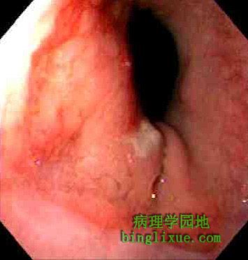 |
如果Barrett黏膜分布在正常的鳞状上皮与柱状上皮交界处上方不超过2厘米,称为短段 Barrett食管。 If the area of Barrett mucosa extends less than 2 cm above the normal squamocolumnar junction, then the condition is called "short segment" Barrett esophagus. |
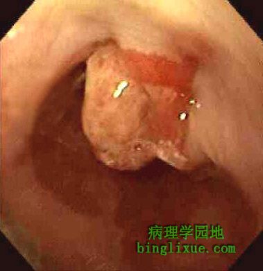 |
胃镜显示食管下段。暗红色碎片状黏膜提示Barrett食管。水螅样的胞块经活检证明是中分化腺癌。病人有30年胃食管返流病史。 This is the view of the lower esophagus seen on endoscopy. Note the areas of dark red friable mucosa representing Barrett esophagus. Note the polypoid mass which on biopsy proved to be a moderately differentiated adenocarcinoma. This patient had a 30 year history of poorly controlled gastroesophageal reflux disease. |
 |
食管下末段(尸解时已经把食管从里面翻出来)线状排列着深兰色的黏膜下层较宽的纹理称为静脉曲张。门静脉高压病人中(慢性酒精性小结节性肝硬化),食管黏膜下血管扩张(形成静脉曲张)。静脉曲张有出血倾向。 At the lower end of the esophagus (which has been turned inside out at autopsy) are linear dark blue submucosal dilated veins known as varices. In patients with portal hypertension (usually micronodular cirrhosis from chronic alcoholism), the submucosal esophageal veins become dilated (form varices). These varices are prone to bleed. |
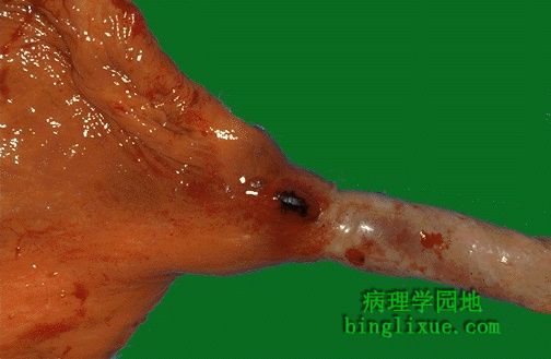 |
胃食管下段静脉曲张,出血使其呈模糊的暗红色。(食管已被翻过来。)静脉丛也包括胃上部血管,但主要是食管静脉丛,因此,此处的出血称为食管静脉曲张性出血。 Here is another varix near the gastroesophageal junction that is dark red black because it has been bleeding. (The esophagus has been turned inside out.) The plexus of veins also involves some of the upper stomach, but it is generically called the esophageal plexus of veins and, hence, bleeding here is termed esophageal variceal bleeding. |
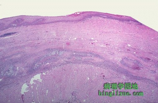 |
鳞状上皮下是被拉长的炎性静脉曲张。静脉曲张出血量大且难以控制。 Below the squamous mucosa is an elongated, inflamed varix. Variceal bleeding can be massive and difficult to control. |
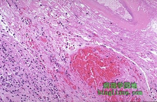 |
食管曲张静脉破裂处见炎症和出血。 Inflammation and hemorrhage is seen here in the region of a ruptured varix of the esophagus. |
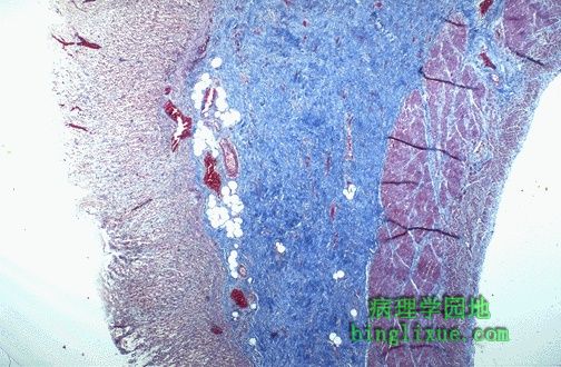 |
系统性硬化(硬皮病)病人食管蠕动障碍,因为黏膜下层纤维化。常常发生在食管,但也可以发生在上消化道任何地方。图示胃粘膜下层广泛纤维化,三色染色呈蓝色。 Esophageal motility problems can occur in patients with progressive systemic sclerosis (scleroderma) because the submucosa becomes fibrotic. This occurs most often in the esophagus, but may also be seen elsewhere in the GI tract. Here in the stomach, a trichrome stain demonstrates a blue submucosa because of the extensive fibrosis. |
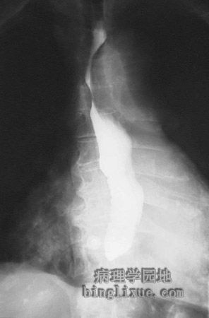 |
X线显示食管下段明显狭窄,与狭窄食管上部相比有明显的差异。这种狭窄见于硬皮病、反流性食管炎、食管癌。 This radiograph taken following barium swallow demontrates a stricture in the lower esophagus, with pooling of the contrast above the point of stricture. Such stricture may complicate conditions such as scleroderma, gastroesophageal reflux disease, or carcinoma. |