
 |
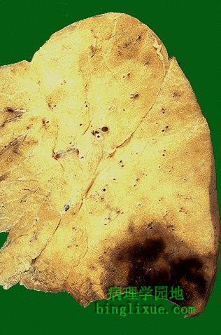
中等大小血栓引起的肺动脉栓塞导致的大面积梗死,梗死灶周围先发生机化。当然也有可能有多个小血栓,但不引起猝死,因为没有堵塞大的肺动脉分支,也就不引起梗死。然而,如果存在大量小血栓,尤其是同时进入肺组织,就会共同堵塞很多小动脉,引起肺高血压。 Here is a larger area of infarction produced by a medium-sized thromboembolus to the lung. This infarction has begun to organize at the margins.It is also possible to have multiple small pulmonary thromboemboli that do not cause sudden death and do not occlude a large enough branch of pulmonary artery to cause infarction. However, if there are lots of small emboli, particularly if they are showered to the lungs over a period of time, then they collectively may block enough small arteries to produce pulmonary hypertension. |
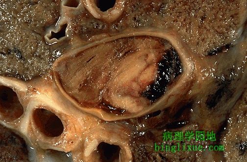 |
肺动脉主干内的血栓栓子近距离外观显示分层结构,这是骨盆或下肢大静脉血栓的特征表现。 A closer view of a thromboembolus filling a main pulmonary artery reveals a layered appearance, typical of a thrombus that formed in a large vein of the pelvis or lower extremity. |
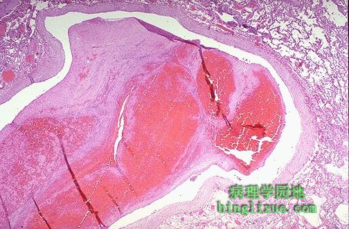 |
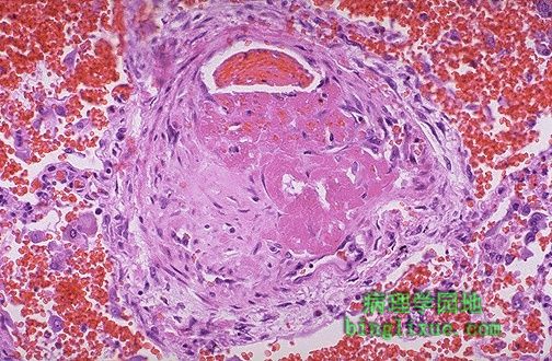
肺动脉大分支内的肺血栓栓子。淡红色及红色的指状突区域形成血栓的特征Zahn线。这些线代表数层细胞,包括红细胞层,血小板层及纤维蛋白层,它们以血栓的形式存在于血管内。 This is the microscopic appearance of a pulmonary thromboembolus in a large pulmonary artery. There are interdigitating areas of pale pink and red that form the "lines of Zahn" characteristic for a thrombus. These lines represent layers of red cells, platelets, and fibrin which are layed down in the vessel as the thrombus forms. |
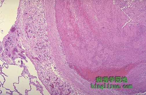 |
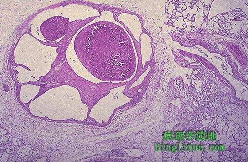
肺动脉栓子,如果病人幸存,将机化和溶解。 Here a thromboembolus is packed into a pulmonary artery. Over time, if the patient survives, the thromboembolus will undergo organization and dissolution. |
 |
小的外周肺动脉血栓栓子。这样的小栓子一般对机体的影响很小,只有众多的小栓子突然或者在一段时间内进入肺循环,才会造成严重影响,可能导致肺高血压。 Here is a small peripheral pumonary artery thromboembolus. Such a small PE such as this one would probably not be noticed or cause problems unless there were many of them showered into the pulmonary circulation at once or over a period of time. This could lead to pulmonary hypertension |
 |
穿过肺动脉分支的结缔组织纤维带显示远端肺栓塞栓子的结构。如果此过程累及许多肺动脉,就可能导致肺动脉高压。 The fibrous bands of connective tissue across this branch of pulmonary artery indicate organization of a remote pulmonary thromboembolus. If many pulmonary arteries are involved by this process, pulmonary hypertension could result. |
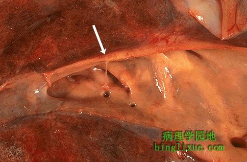 |
白色箭头之下可见外周肺动脉上的一个纤维带,是远端肺动脉栓塞栓子机化形成的。注意右边肺动脉内膜上的动脉粥样斑块,表现出栓塞的指征肺动脉高压。 Below the white arrow can be seen a fibrous band in a peripheral pulmonary artery from a remote organized pulmonary thromboembolus. Note that the atheromatous plaques of the pulmonar artery intima at the right are indicative of the effect of such embolization--pulmonary hypertension. |
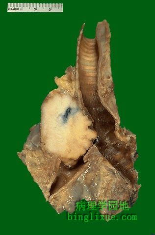 |
发生于肺中央(与绝大多数鳞状细胞癌一样)的鳞状细胞癌,刚好阻挡右主支气管。肿瘤质地坚韧,切面呈灰白色。 This is a squamous cell carcinoma of the lung that is arising centrally in the lung (as most squamous cell carcinomas do). It is obstructing the right main bronchus. The neoplasm is very firm and has a pale white to tan cut surface. |
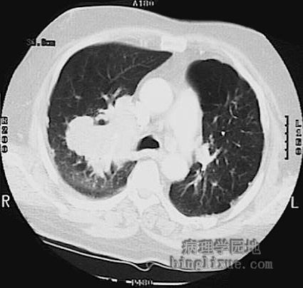 |
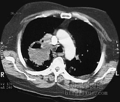
胸部CT显示(上下两图)右上肺叶鳞状细胞癌,累及右主支气管、浸润纵隔、破坏肺门淋巴结。 These chest CT scan views above and below demonstrates a large squamous cell carcinoma of the right upper lobe that extends around the right main bronchus and also invades into the mediastinum and involves hilar lymph nodes. |
 |