
 |
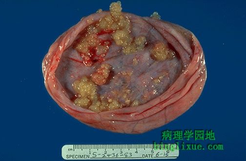
This is a papillary serous cystadenocarcinoma. Note the many papillations on the inner surface. Between benign cystadenomas and malignant cystadenocarcinomas lies the grey zone of "borderline" lesions that are not clearly malignant, but are treated as though they could be. 图示乳头状浆液性囊腺癌。 内表面可见许多乳头状突起。良性的囊腺瘤和恶性的囊腺癌的病变界限并不十分分明。虽然病变可能并不是明显的恶性,但通常按恶性对待。 |
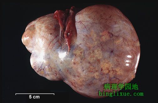 |
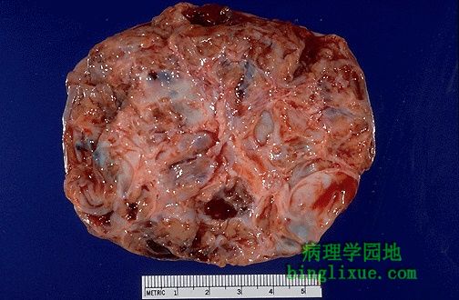
This ovarian papillary cystadenocarcinoma is mostly composed of solid tissue and has invaded outside of the ovary, with papillations seen over the surface. Because there are no early signs or symptoms with masses in the ovary, many of these ovarian tumors have metastasized by the time they are detected with abdominal enlargement. These neoplasms characteristically spread by "seeding" along peritoneal surfaces. 乳头状浆液性囊腺癌是由大量的实体组织构成,并已侵袭至卵巢外。表面可见乳头状突起。由于早期卵巢肿块没有相应的症状和体征,因此当由于腹部增大而检查出来时,许多已经转移了。转移特点是沿腹膜表面的种植性转移。 |
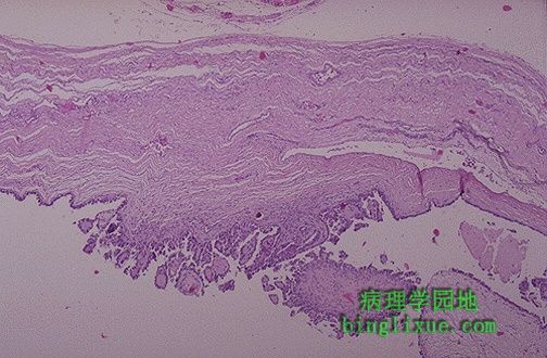 |
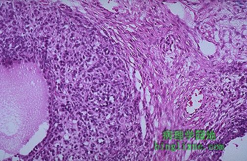
Microscopically, a borderline serous cystadenoma is seen here with papillary projections of epithelium extending into the lumen of the tumor. There is no invasion of the stroma or capsule. |
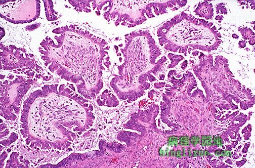 |
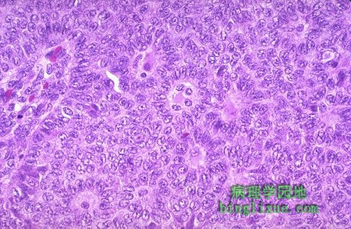
Here is a serous cystadenocarcinoma in which there is more pronounced papillary growth with more hyperchromatic cells. 图示浆液囊腺癌,其中有许多由大量深染细胞形成的明显的乳头状突起。 |
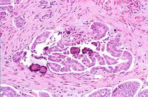 |
Ovarian papillary serous cystadenocarcinomas may contain small concretions called psammomma bodies, seen here as purplish rounded and laminated objects. They are essentially just a form of dystrophic calcification in neoplasms 卵巢乳头状浆液性囊腺癌可包含小的砂粒体,如图所见的略带紫色的圆片状的物质,由肿瘤的营养不良性钙化形成。 |
 |
This is a granulosa cell tumor of ovary with a variegated cut surface. These tumors are derived from the ovarian stroma and often have a component of thecoma. They are often hormonally active and can produce large amounts of estrogen such that the patient may initially present with bleeding from endometrial hyperplasia. 卵巢颗粒细胞瘤,有杂色切面,源于卵巢间质。常常激素分泌活跃,产生大量的雌激素,使得患者最初常有子宫内膜增生所致的出血。 |
 |
Microscopically, the granulosa cell tumor attempts to form structures that resemble primitive follicles, as seen at the left. Most of these tumors are histologically benign, but some are malignant. 镜下,颗粒细胞瘤趋向于形成类似于原始的滤泡的结构,如图左示。大多数在组织学上呈良性,但一些是恶性的。 |
 |
At higher magnification, an ovarian granulosa cell tumor has nests of cells which are forming primitive follicles. 高倍镜示:卵巢颗粒细胞瘤有形成原始滤泡的细胞巢。 |
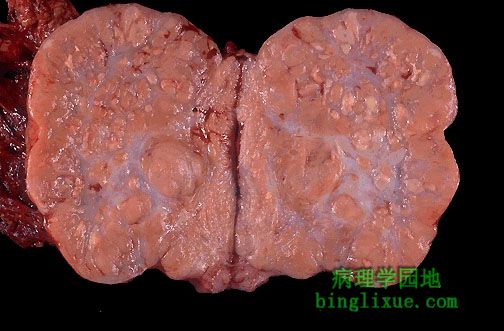 |
This is an ovarian dysgerminoma that has been sectioned into two halves. Note the pale brown appearance of the parenchyma, along with some central collagenous scar. The gross and microscopic appearance of an ovarian dysgerminoma is essentially the same as a seminoma of the testis in a male. 图示:卵巢无性细胞瘤,已被剖开。实质呈浅褐色外观,中央有一些胶原块。卵巢的无性细胞瘤肉眼和显微镜下表现基本上与男性睾丸的精原细胞瘤一致。 |
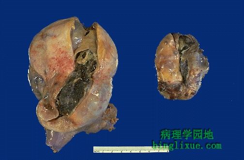 |
Here are bilateral mature cystic teratomas of the ovaries. These are a form of ovarian germ cell tumor. Histologically, a variety of mature tissue elements may be found. These tumors are often called "dermoid cysts" because they are mostly cystic. 双侧卵巢成熟囊性畸胎瘤,是来源于生殖细胞的肿瘤。组织学上,可见多种成熟组织成分。这些肿瘤常常被称为“皮样囊肿”,因形成囊状。 |