
 |
This is normal secretory phase endometrium. Note the larger tortuous glands with secretions. The secretory phase follows a set 14 day course leading to either implantation of a fertilized ovum or menstruation. 图示正常分泌期子宫内膜。腺体长而屈曲。分泌期约经历14日左右,此期间卵子若受精,就形成受精卵并着床。若未受精,就进入下一个月经周期。 |
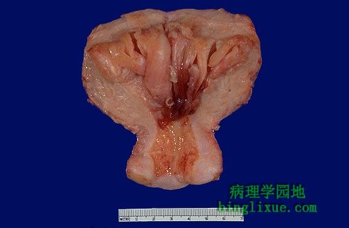 |
The endometrial cavity is opened to reveal lush fronds of hyperplastic endometrium. Endometrial hyperplasia usually results with conditions of prolonged estrogen excess and can lead to metrorrhagia (uterine bleeding at irregular intervals), menorrhagia (excessive bleeding with menstrual periods), or menometrorrhagia. 图示子宫内膜增生,常由于雌激素过多引起,导致子宫不规则出血(周期不规则),月经过多(周期规则,经量过多),子宫不规则过多出血(周期不规则,血量过多)。
|
 |
This uterus has been opened anteriorly through cervix and into the endometrial cavity. High in the fundus and projecting into the endometrial cavity is a small endometrial polyp. Such benign polyps may cause uterine bleeding. 自宫颈至子宫腔,在前部打开,可见基底部有小的子宫内膜息肉伸向宫腔。这种良性息肉会导致子宫出血。 |
 |
This is endometrial cystic hyperplasia in which the amount of endometrium is abnormally increased and not cycling as it should. The glands are enlarged and irregular with columnar cells that have some atypia. Simple endometrial hyperplasias can cause bleeding, but are not thought to be premalignant. However, adenomatous hyperplasia is premalignant. 图示子宫内膜囊性增生,大量子宫内膜呈不规则增生,腺体不规则增大,柱状上皮有一定的异型性。单纯型子宫内膜增生可导致出血,但不至于成为癌前病变。但是腺瘤状内膜增生可进展为癌。 |
 |
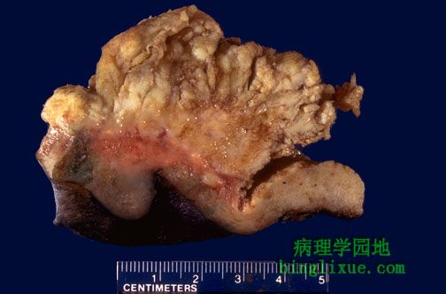
This uterus is not enlarged, but there is an irregular mass in the upper fundus that proved to be endometrial adenocarcinoma on biopsy. Such carcinomas are more likely to occur in postmenopausal women. Thus, any postmenopausal bleeding should make you suspect that this lesion may be present.
图示子宫无增大,基底部有一不规则肿物,已取活检确定为子宫内膜癌。子宫内膜癌多发生于绝经后妇女,因此绝经后出血应该警惕内膜癌的可能。 |
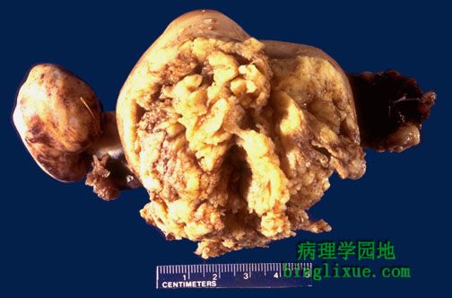 |
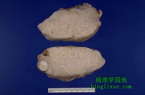
This adenocarcinoma of the endometrium is more obvious. Irregular masses of white tumor are seen over the surface of this uterus that has been opened anteriorly. The cervix is at the bottom of the picture. This enlarged uterus was no doubt palpable on physical examination. Such a neoplasm often present with abnormal bleeding. 图示子宫内膜腺癌。自前部打开的子宫可见不规则肿物呈白色。图下方示宫颈。在检查中可触及增大的子宫。此种肿瘤常造成不规则出血。
|
 |
The endometrial adenocarcinoma is present on the lumenal surface of this cross section of uterus. Note that the neoplasm is superficially invasive. The cervix is at the right. 图示子宫内膜腔面纵切面的子宫内膜腺癌,可见其向下浸润。右示宫颈。 |
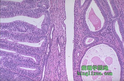 |
The endometrial adenocarcinoma in the polyp at the left is moderately differentiated, as a glandular structure can still be discerned. Note the hyperchromatism and pleomorphism of the cells, compared to the underlying endometrium with cystic atrophy at the right. 图左 示中度分化的子宫内膜腺癌,可见腺状结构,与右侧囊性萎缩的子宫内膜相比,注意细胞染色深及多型性。 |
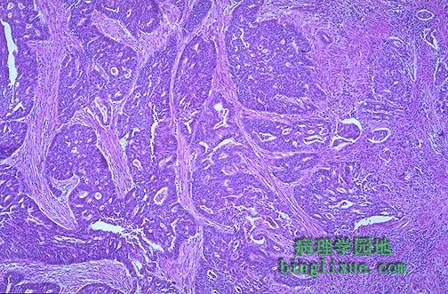 |
This is endometrial adenocarcinoma which can be seen invading into the smooth muscle bundles of the myometrial wall of the uterus. This neoplasm has a higher stage than a neoplasm that is just confined to the endometrium or is superficially invasive. 图示子宫内膜腺癌侵入肌层,此为高分期的肿瘤。 |
 |
The thickened and spongy appearing myometrial wall of this sectioned uterus is typical of adenomyosis. There is also a small white leiomyoma at the lower left. 在子宫肌壁中出现粗厚和海绵状的团块,这是子宫腺肌病的典型表现。图左下方示一体积较小的白色的平滑肌瘤。 |