
 |
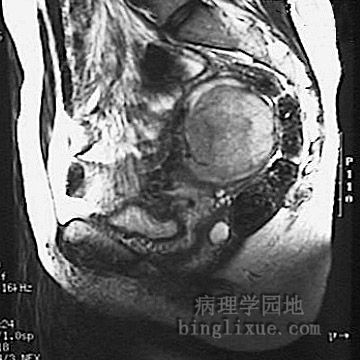
This magnetic resonance imaging (MRI) scan of the pelvis in sagittal view demonstrates a large discreet leiomyoma of the upper posterior myometrium, seen just anterior to the rectosigmoid colon. MRI示盆腔矢状面,于后上方子宫肌层可见一体积大,界限清楚的子宫肌瘤,后方紧邻乙状结肠。
|
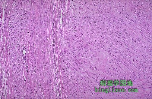 |
Here is the microscopic appearance of a benign leiomyoma. Normal myometrium is at the left, and the neoplasm is well-differentiated so that the leiomyoma at the right hardly appears different. Bundles of smooth muscle are interlacing in the tumor mass. 镜下示良性平滑肌瘤。左侧为正常子宫平滑肌,右侧为高分化的平滑肌瘤,与正常者几乎无异,平滑肌瘤中可见平滑肌成编织状。 |
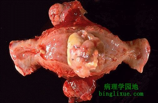 |
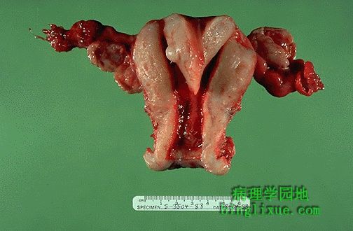
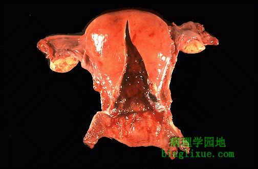
This is a leiomyosarcoma protruding from myometrium into the endometrial cavity of this uterus that has been opened laterally so that the halves of the cervix appear at right and left. Fallopian tubes and ovaries project from top and bottom. The irregular nature of this mass suggests that is not just an ordinary leiomyoma. 示自肌层突向宫腔的平滑肌肉瘤。标本自中央剖开,图片左侧右侧为宫颈,左右侧输卵管和卵巢位于图片的上下方。此肿瘤从大体上显示出与一般的平滑肌瘤有所差别。 |
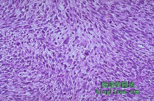 |
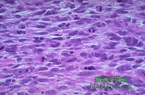
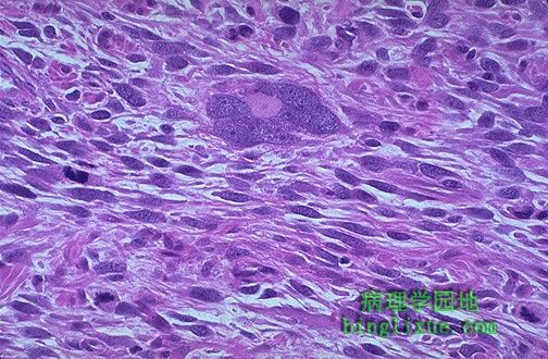
Here is the microscopic appearance of a leiomyosarcoma. It is much more cellular and the cells have much more pleomorphism and hyperchromatism than the benign leiomyoma. An irregular mitosis is seen in the center. 镜下示平滑肌肉瘤的特点。与良性的平滑肌瘤相比,其细胞丰富,细胞异型性明显,深染。中央可见病理性核分裂象。 |
 |
As with sarcomas in general, leiomyosarcomas have spindle cells. Several mitoses are seen here, just in this one high power field. 正如肉瘤的一般特点,平滑肌肉瘤中可见梭形细胞,仅一个高倍视野中就可见多个病理性核分裂象。 |
 |
Sarcomas, including leiomyosarcomas, often have very large bizarre giant cells along with the spindle cells. A couple of mitotic figures appear at the left and lower left. 包括平滑肌肉瘤在内的肉瘤中常可见到形状奇异的巨细胞,分布于梭性细胞之间。图左侧及左下方可见核分裂象。 |
 |
Here is a bifid (septate) uterus. Sometimes even the cervix and/or vagina may be double as well. This is of no major consequence except that in pregnancy a bifid uterus may not enlarge normally and lead to fetal loss, or a normal vaginal delivery may not be possible with a double cervix or vagina. Note that there is also a small intramural leiomyoma on the septum at the left. 图示双房子宫(又称有隔子宫),是子宫畸形的一种,有时也会形成双宫颈,双阴道。此种畸形不会造成严重的临床症状,但是在怀孕的时候,双房子宫不能随着胎儿的生长发育而伸展,因而造成流产。另外如果合并双宫颈、双阴道,胎儿不可能经阴道分娩。注意子宫中隔左侧还可见一小的肌壁间平滑肌瘤。 |
 |
Ovulation releases an egg from an ovarian follicle. The egg is swept into the fallopian tube and begins to descend. Spermatozoa (millions are represented here by one) begin ascending. Fertilization of the egg by a single sperm occurs in the ampullary portion of the fallopian tube about a day after ovulation. The fertilized egg begins to develop into the blastocyt on descent into the endometrial cavity, where implantation occurs on the wall of the fundus about a week after ovulation. 图示排卵,受精及受精卵的发育,输送和着床。排卵是指卵泡将卵细胞和它周围的一些细胞排出,卵子排除后,经输卵管伞部拾拣,开始沿着输卵管下行。数百万的精子则向上运行,在输卵管的壶腹部,精子和卵子结合,称为受精,整个受精过程大约需要24小时。受精卵进一步分裂成为桑椹胚,并向宫腔方向移动,受精后大概1周左右受精卵种植于宫腔底部。 |
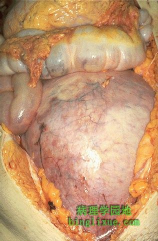 |
Here is the size of the uterus in the third trimester. Note how it displaces the bowel superiorly and fills the lower abdomen. (This unfortunate woman died accidentally, and the baby died too). 图示晚期妊娠子宫的体积。注意子宫都达到横结肠的位置,并填充满下腹部。(这位不幸的孕妇死于意外,胎儿也死于母体内。) |
 |
This is a normal postpartum uterus 5 days following delivery. Note how quickly the uterus is returning to its normal non-pregnant size. 图示正常分娩5天后的子宫。产后10天后子宫即可降至骨盆腔内,腹部检查扪不到宫底。因此子宫复旧的速度是很快的。 |