
 |
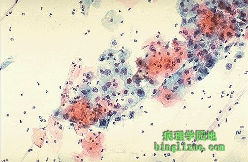
This is a Pap smear. The cytologic features of normal squamous epithelial cells can be seen at the center top and bottom, with orange to pale blue plate-like squamous cells that have small pyknotic nuclei. The dysplastic cells in the center extending to upper right are smaller overall with darker, more irregular nuclei. 这是一张巴氏涂片。图中上部及下部示正常鳞状上皮细胞特征,鳞状细胞呈红色、淡蓝色,核小而固缩。图中央至右上示不典型增生细胞,细胞体积小,核染色深,不规则.
|
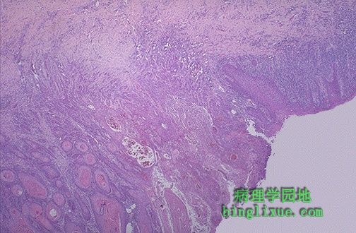 |
This is why you do Pap smears--to prevent invasive squamous cell carcinomas from occurring. With Pap smears, pre-neoplastic and neoplastic cervical lesions can be detected when small and treated. Nests of squamous cell carcinoma have invaded underlying stroma at the center and left. 巴氏涂片可以使宫颈鳞癌以及其癌前病变得到较早的诊断和治疗。图中、左示鳞状细胞癌的间质浸润。 备注:宫颈涂片检查是目前宫颈癌普查常规使用的方法。宫颈涂片检查是取宫颈脱落细胞涂片,然后酒精固定,采用巴氏染色法观察细胞形态,以初步了解有无癌存在。巴氏Ⅰ级为正常细胞涂片,巴氏Ⅱ级为炎症细胞;Ⅲ级代表可疑癌;Ⅳ级代表高度可疑癌;Ⅴ级肯定为癌
|
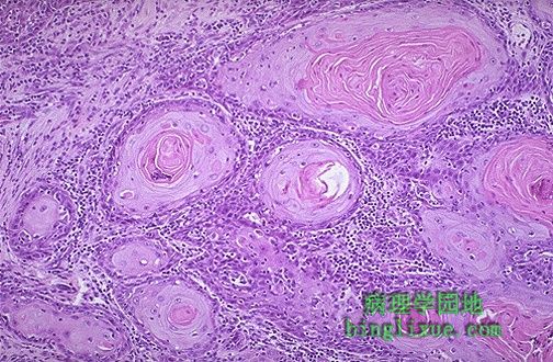 |
At high magnification, nests of neoplastic squamous cells are invaded through a chronically inflamed stroma. This cancer is well- differentiated, as evidenced by keratin pearls. However, most cervical squamous carcinomas are non-keratinizing. 高倍镜下示鳞癌浸润到间质,间质中有慢性炎症。鳞癌分化程度高,可见角化珠。但大部分宫颈鳞癌为非角化型。 |
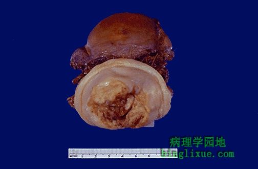 |
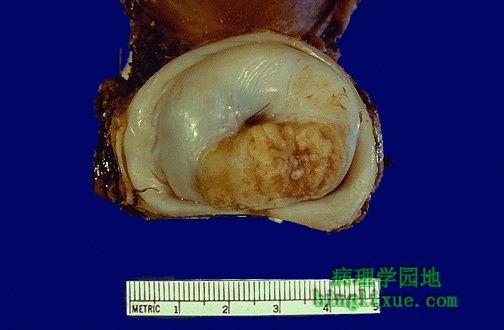
This is the gross appearance of a cervical squamous cell carcinoma that is still limited to the cervix (stage I). The tumor is a fungating red to tan to yellow mass. 图示宫颈鳞癌(I期)的大体标本,病变局限于宫颈。肿瘤呈菌样生长,黄褐色。 |
 |
Here is another cervical squamous cell carcinoma. Note the IUD string protruding from the cervix. This implies that someone could have done a Pap smear when it was inserted. There is a natural history of progression of dysplasia to carcinoma, so don't leave dysplasias alone. 图示另例宫颈鳞癌的大体观。注意宫颈处突出的宫内节育器,这表示在植入前做过巴氏涂片。 从宫颈不典型增生到癌是一个逐渐发展的自然病程,因此不能忽略不典型增生的病变。 |
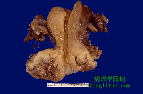 |
This is a larger cervical squamous cell carcinoma which spread to the vagina. A total abdominal hysterectomy with bilateral salpingo-oopherectomy (TAH-BSO) was performed. 图示宫颈鳞状细胞癌,已扩散至阴道。已行经腹子宫加双侧输卵管、卵巢切除术(TAH-BSO) |
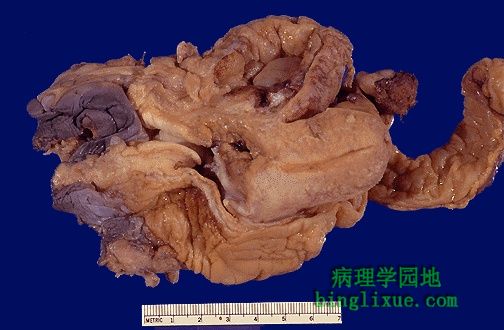 |
This is a pelvic exenteration done for stage IV cervical carcinoma. At the left, dark vulvar skin leads to vagina and to cervix in the center, where an irregular tan tumor mass is seen infiltrating upward to the bladder. A slit-like endometrial cavity is surrounded by myometrium at the mid-right. The rectum and sigmoid colon are at the bottom extending to the right. 图示IV期宫颈肿瘤,行盆腔清除术。图左示外阴(皮肤色深)、阴道,图中示宫颈肿瘤,一不规则褐色肿物,向下浸润至膀胱。图中偏右示裂缝状的子宫内膜腔,由子宫肌层包绕。图下方至右方示示直肠、乙状结肠。 |
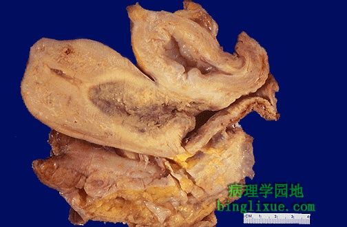 |
This is another pelvic exenteration for cervical squamous cell carcinoma. The irregular grey-brown tumor extends toward bladder and up into the uterus. 另例宫颈鳞状细胞癌,行盆腔清除术。不规则肿物呈灰褐色,已浸润至膀胱和子宫.
|
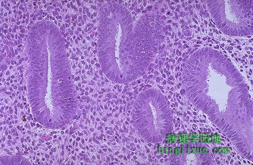 |
This is the microscopic appearance of normal proliferative endometrium in the menstrual cycle. The proliferative phase is the variable part of the cycle. In this phase, tubular glands with columnar cells and surrounding dense stroma are proliferating to build up the endometrium following shedding with previous menstruation. 镜下显示正常增生期子宫内膜。在正常月经周期中的增生期,随着上次月经周期内膜的脱落,子宫内膜腺体柱状上皮和间质细胞呈增生状态。 |
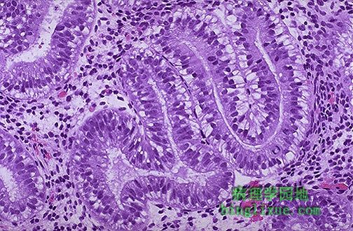 |
Here is early secretory endometrium. The appearance with prominent subnuclear vacuoles in cells forming the glands is consistent with post-ovulatory day 2. The histologic changes following ovulation are quite constant over the 14 days to menstruation and can be utilized to date the endometrium. 图示早期分泌期子宫内膜。腺上皮细胞出现明显的核下空泡,图像符合排卵后第二天子宫内膜表现。排卵后14日子宫内膜都呈现此种组织学形态。
|