
 |
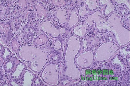
女性乳腺在妊娠期间会经历乳腺小叶增生以至于以对分娩后的哺乳。图中可见充满粉红色分泌物的乳腺小叶。乳腺组织学上类似于汗腺,以出芽方式分泌。 The female breast during pregnancy undergoes lobular hypertrophy so that following birth lactation can occur. Seen here are lobules filled with pink secretions. The breast, which histologically is a modified sweat gland, secretes by budding off of portions of cell cytoplasm. |
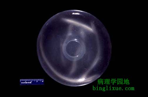 |
图示隆胸术所用硅酮。手术可使乳腺形状再塑,体积增大。保持自然形状和触感是硅酮的特点。 Here is a silicone breast implant. These implants are used for breast augmentation and for breast reconstruction following surgery. The silicone provides for a natural shape and feel. |
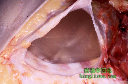 |
图示乳腺硅酮植入物的薄结缔组织被膜。值得注意的是左上方是叠压的皮肤与脂肪组织,在植入物之下靠右边的是胸壁。这是典型的圆滑而不变形的被膜。 The thin connective tissue capsule around a silicone breast implant is shown here. Note the overlying skin and adipose tissue at the upper left with the chest wall below the implant and to the right. This is a typical capsule that is pliable and non-deforming. |
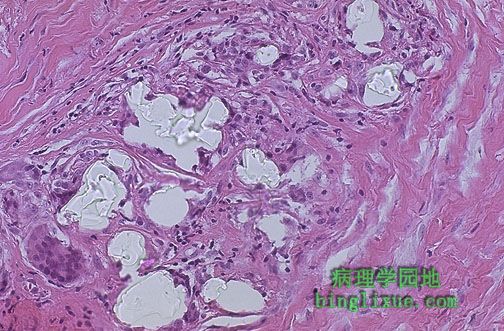 |
乳腺硅酮植入物纤维被膜镜检时常可显示出屈光性硅酮材料,起因于这种材料渐渐漏出。可见异物肉芽肿性反应。并未显示其与系统疾病如自身免疫病相关。乳腺植入物周围的纤维化可能在某些妇女中引起变形与疼痛。被膜破裂不常见。 Microscopic examination of the fibrous capsule from a silicone breast implant will often reveal the refractile silicone material as shown here, because this material gradually leaks out. There is a foreign body granulomatous response. This has not been shown to be associated with systemic disease, such as autoimmune disease. The fibrosis around a breast implant may produce deformity and pain in some women. Rupture is uncommon. |
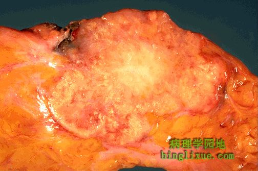 |
不规则块状病变是乳腺浸润性导管癌。中央坚硬(硬癌)且因纤维结缔增生呈白色。肿瘤淡黄*色坏死区向周围乳腺组织浸润。这种肿瘤通常坚硬不活动。 The irregular mass lesion seen here is an infiltrating ductal carcinoma of breast. The center is very firm (scirrhous) and white because of the desmoplasia. There are areas of yellowish necrosis in the portions of neoplasm infiltrating into the surrounding breast. Such tumors appear very firm and non-mobile on physical exam. |
 |
乳腺X线显示大肿块样病变,经针吸细胞学检查证实为癌。 This mammogram demonstrates a mass lesion that was a carcinoma on fine needle aspiration (FNA) cytologic examination. |
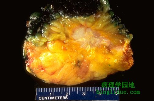 |
乳腺活组织检查显示为癌。值得注意的是不规则的边缘与多样的切面。此小癌块是经乳腺X射线照相发现的。肿块边缘被墨水染成绿色,目的是在组织学切片做成后辅助判断癌浸润到边缘与否。 This breast biopsy demonstrates a carcinoma. Note the irregular margins and varied cut surface. This small cancer was found by mammography. The margins of the specimen have been inked with green dye following removal to assist in determining whether cancer extends to the margins once histologic sections are made. |
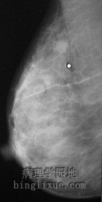 |
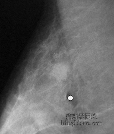
乳腺X线显示白点标记的肿瘤性病变,病人在标记区有触痛。活检证实为乳腺浸润性导管癌。 This mammogram demonstrates a lesion consistent with a neoplasm in the upper portion above and just to the left of the white dot marking the point the patient felt some pain on palpation. On biopsy, this was an infiltrating ductal carcinoma. |
 |
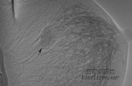
放大的乳腺X线片,显示病变周围钙化。 A higher magnification of this lesion, demonstrating tiny peripheral calcifications, is seen below. |
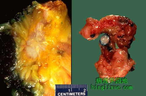 |
左边典型的浸润性导管癌与右边良性纤维腺瘤的肉眼特征的近距离比较。 Here is a side by side comparison of the gross characteristics of a classic infiltrating ductal carcinoma on the left and a benign fibroadenoma on the right. |