
 |
The normal microscopic appearance of female breast tissue is shown here. There is a larger duct to the right and lobules to the left. A collagenous stroma extends between the structures. A variable amount of adipose tissue can be admixed with these elements. 正常女性乳腺显微结构。左侧可见乳腺小叶,右侧可见乳导管,它们之间是胶原基质,不定量的脂肪组织分布于这些结构之间。 |
 |
At high magnification, the appearance of a normal breast acinus is shown here. Note the epithelial cells lining the lumen demonstrate apocrine secretion with snouting, or cytoplasmic extrusions, into the lumen. A layer of myoepithelial cells, some of which are slightly vacuolated, is seen just around the outside of the acinus. 高倍镜显示正常乳腺腺泡。值得注意的是排成管腔的上皮细胞显示了分泌物进入管腔的顶浆分泌或细胞质分泌。一层肌上皮细胞,部分呈轻微空泡状,正好围绕在腺泡的外围。 |
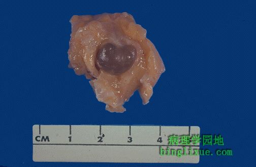 |
This is the gross appearance of fibrocystic changes in the breast. A 1.5 cm cyst is noted here. This can lead to palpation of an ill-defined "lump" in the breast. Sometimes, fibrocystic changes produce a more diffusely lumpy breast. 乳腺纤维囊性变大体外观。可见一1.5cm的囊。乳腺可触诊到不明原因的“肿块”。有时纤维囊性变导致乳腺更多的肿块出现。 |
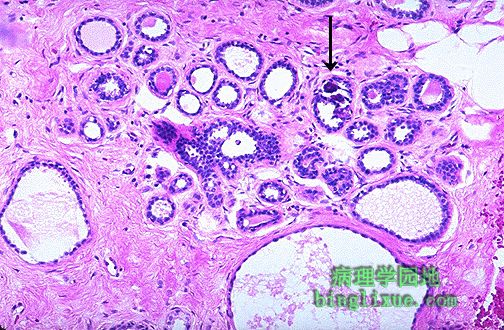 |
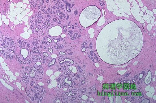
This is the histologic appearance of fibrocystic changes in breast. There are cystically dilated ducts, areas of lobules that are laced with abundant fibrous connective tissue (sclerosing adenosis), and stromal fibrosis. There is even a small area of microcalcification seen just to the upper right of center. No atypical changes are seen here. 这是乳腺纤维囊性变的组织学外观。可见到囊性扩张导管、小叶区伴大量纤维结缔组织(硬化性腺病)增生、间质纤维化。靠近中心的右上部可见一小钙化灶。未见非典型增生。 |
 |
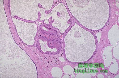
Another example of microscopic fibrocystic changes of the breast are shown here. Fibrocystic changes account for the majority of "breast lumps" that are found in women of reproductive years, particularly between age 30 and menopause. 又一例乳腺纤维囊性变。大多数发现于生育期妇女尤其是在30岁到绝经期之间的“乳腺肿块”是由纤维囊性变引起的。 |
 |
At low power, the prominent cysts of fibrocystic changes are shown. The cysts are lined by a single epithelial layer of varying height. 低倍镜显示纤维囊性变。囊是由高度不一的单层上皮排列而成。 |
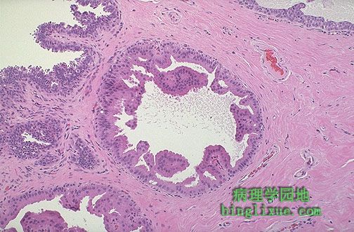 |
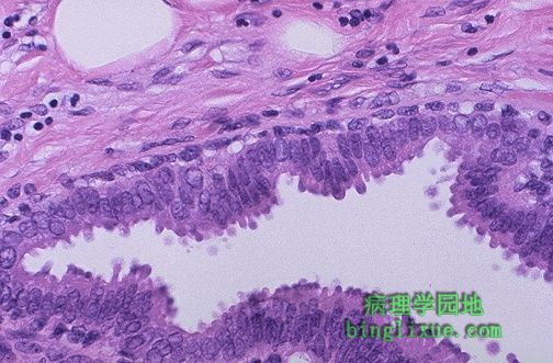
此例乳腺纤维囊性变有排列成囊的细胞的显著顶浆分泌改变。值得注意的是上皮细胞呈现其原本的粉红色高柱状。这种外观是良性的。 There is prominent apocrine change of the cells lining the cysts in this example of fibrocystic changes of breast. Note the tall, pink, columnar nature of the epithelial cells. This appearance is benign. |
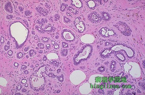 |
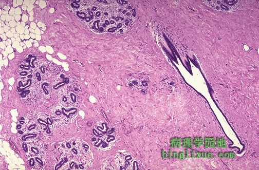
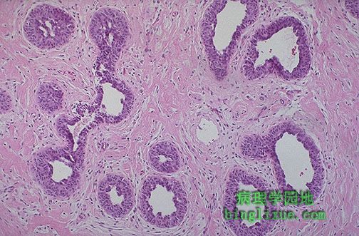
纤维间质中小导管增生显示显著的硬化性腺病,其为纤维囊性变的特征之一。虽然它是良性的,肉眼与X线看到的外观却仿佛是癌,且很难通过冰冻切片将它与癌区分开来。 Prominent sclerosing adenosis, one of the features of fibrocystic changes, is demonstrated by the appearance of a proliferation of small ducts in a fibrous stroma. Although it is benign, the gross and mammographic appearance may mimic carcinoma, and it can be difficult to distinguish from carcinoma on frozen section. |
 |
乳腺X线显示可疑病变可能是癌也可能是硬化性腺病的囊性病变。活体组织检查显示为良性病变。 This mammogram demonstrates a suspicious lesion that could be a carcinoma or just an area of pronounced sclerosis with fibrocystic changes. On biopsy, this was benign. |
 |
乳腺导管显示上皮细胞增生。上皮细胞呈多层,未见异型性。因而只有纤维囊性变,如纤维性增生、囊性变和硬化性腺病,没有增加患癌的风险。 These breast ducts demonstrate epithelial hyperplasia. The epithelial cells are multilayered. There is no atypia. Thus, just as with fibrocystic changes such as fibrosis, cysts, and sclerosing adenosis, there is no increased risk for carcinoma. |