
 |
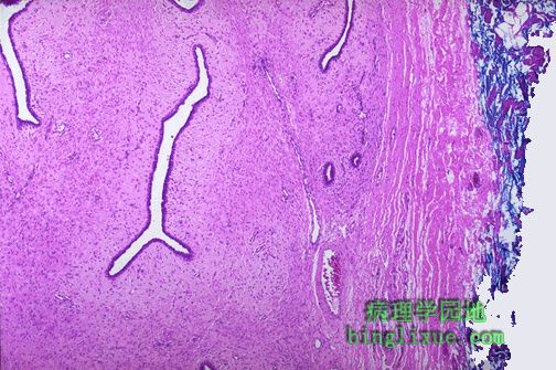
乳腺导管上皮呈旺炽性增生。患乳腺癌的风险轻微增加(为正常的1.5到2倍)。 More florid ductal epithelial hyperplasia of the breast is shown here. There is a slightly increased risk (1.5 to 2 times normal) for breast carcinoma when such changes are present. |
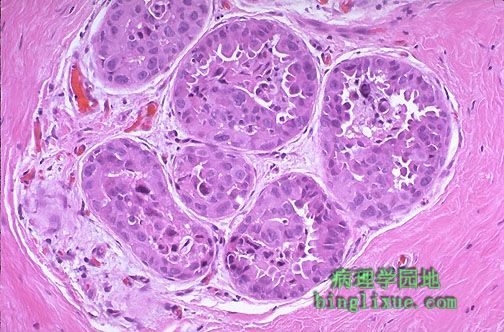 |
乳腺导管上皮细胞非典型增生。非典型上皮细胞增生出现使乳腺癌风险明显增加(是正常值的5倍)。 This is atypical ductal epithelial hyperplasia of the breast. A significantly increased risk (5 times normal) for breast carcinoma occurs with cytologically atypical epithelial hyperplasia. |
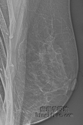 |
乳腺X线显示伴微钙化灶的可疑病变,可能为癌或纤维囊性病变。活体组织检查显示为伴上皮增生的纤维囊性变。 This mammogram demonstrates a suspicious area with microcalcifications that could be a carcinoma or just an area of fibrocystic changes. On biopsy, this lesion had areas of fibrocystic changes with epithelial hyperplasia. |
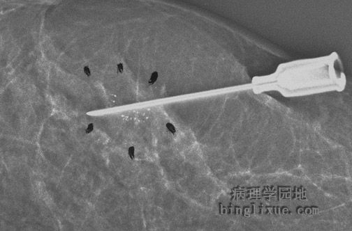 |
乳腺X线显示应用针头定位辅助外科手段切除可疑病变。 The mammogram below demonstrates the use of needle localization to aid the surgeon in excising the suspicious area. |
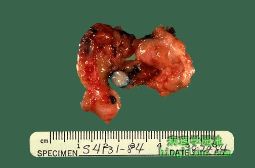 |
乳腺小肿块的外科手术切除标本,肿块边界清楚,质硬有弹性,为纤维腺瘤。纤维腺瘤周围的蓝色染剂是在放射学上准确定位时标记病变的,使外科医生更容易找到小肿块。 Here is a surgical excision of a small mass from the breast. The mass is well-circumscribed. Grossly it felt firm and rubbery. This is a fibroadenoma. The blue dye around the fibroadenoma was used to mark the lesion during needle localization in radiology so that the surgeon could find this small mass. |
 |
纤维腺瘤的显微外观。靠右方受压的乳腺纤维结缔组织形成了肿块被膜。肿瘤由纤维化的间质组成,间质中有着扁平的长导管,导管是由呈良性上皮排列而成。 Here is the microscopic appearance of a fibroadenoma. To the right is compressed breast connective tissue forming a "capsule" to this mass. The neoplasm itself is composed of a fibroblastic stroma in which are located elongated compressed ducts lined by benign appearing epithelium. |
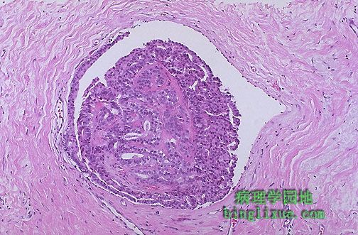 |
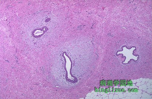
图示乳腺导管中的小的良性导管内乳头状瘤,代表性地出现于乳晕下的一个主要输乳管内。值得注意的是上皮细胞未显示异型性且在乳头状瘤有清晰可见的粉红色胶原间质。导管内乳头状瘤可能产生浆液性或血性乳头溢液,也可能引起某种程度的乳头内陷。 A small benign intraductal papilloma appears here in a breast duct, typically in one of the main lactiferous ducts beneath the areola. Note that the epithelial cells show no atypia and that there is a fine pink collagenous stroma within the papilloma. An intraductal papilloma may be associated with a serous or bloody nipple discharge, or it may cause some nipple retraction. |
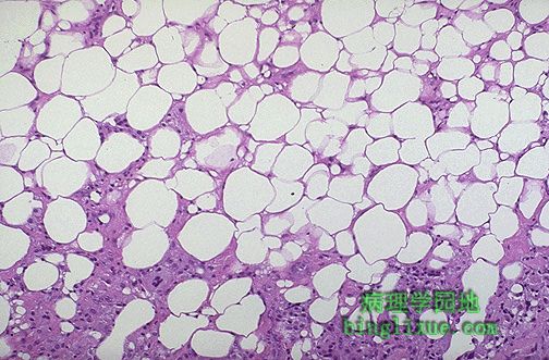 |
乳腺脂肪坏死。最常见的病因是创伤。它可因纤维化成为局限区以致于灰类似于乳腺癌外观。然而在显微镜下,脂肪坏死是由不规则无细胞核的消脂细胞组成,夹杂着粉红色无定形坏死物和炎细胞,包括吞噬了坏死脂肪细胞的异物巨细胞。 This is fat necrosis of the breast. The most common etiology is trauma. It can be a localized, firm area with scarring that can mimic a breast carcinoma. Microscopically, however, fat necrosis consists of irregular steatocytes with no peripheral nuclei and intervening pink amorphous necrotic material and inflammatory cells, including foreign body giant cells responding to the necrotic fat cells. |
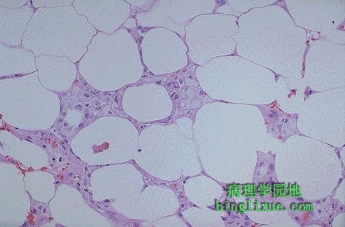 |
脂肪坏死的高倍图中可见到一些充满脂质的巨噬细胞位于坏死的脂肪组织细胞之间。最常见的病因是创伤,但乳腺脂肪坏死也有因外科手术和放射治疗引起的。 In this view of fat necrosis at high magnification, some lipid-laden macrophages are seen between the necrotic adipose tissue cells. The most common etiology is trauma, but fat necrosis of the breast can also occur with surgery and radiation therapy. |
 |
男性有少量的乳腺组织,但它仅有一些导管组成,纤维间质中没有小叶。它可能是单侧的也可能是双侧的。有时它可能出现于青春期或者随年龄增长而出现。男性乳腺发育可由肝硬化、睾丸间质细胞瘤或药物引起。可见到导管上皮细胞增生或如这里所见有明显的导管水肿。 Males have a small amount of breast tissue, but it consists of just a few ducts, without lobules, in a fibrous stroma. It may be unilateral or bilateral. Sometimes it can occur at puberty or sometimes with aging. Gynecomastia may occur with cirrhosis of liver, Leydig cell tumors of testis, or with drugs. There can be ductal epithelial hyperplasia, or prominent periductular edema as seen here. |