
 |
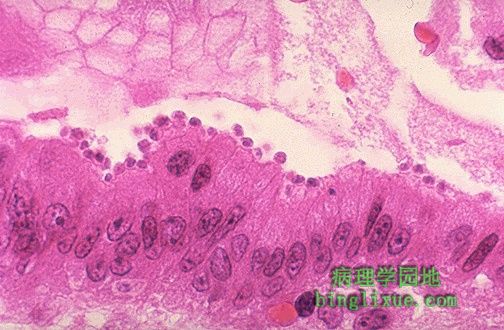
沿着小肠上皮刷状边缘排列的呈蓝色的病原体是隐孢子虫 。隐孢子虫感染使免疫缺陷病人腹泻。 The little blue organisms lined up along the brush border of the small intestinal epithelium are Cryptosporidia. This infection causes diarrhea in immunocompromised hosts. |
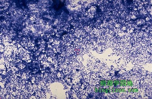 |
隐孢子虫感染最好采用粪便检测。采用抗酸染色在中央可见3个孢囊。 Cryptosporidium infection is best diagnosed by stool exam. Three cysts are seen in the center with this acid fast stain. |
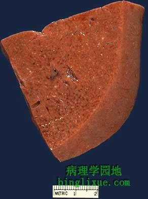 |
许多真菌感染都能形成肉芽肿,免疫缺陷病人免疫反应很差,肉芽肿很少形成,或根本不会形成。肝脏标本中黄褐色部分为肉芽肿,该病人患组织胞浆菌病。 Many fungal infections can produce a granulomatous pattern. In immunocompromised hosts, the immune response is often poor, so granulomas are poorly formed, if at all. This portion of liver demonstrates some pinpoint yellow-tan granulomas in a patient with disseminated histoplasmosis. |
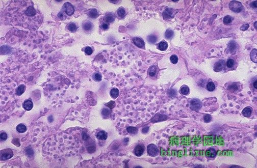 |
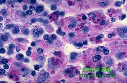
荚膜组织胞浆菌感染。注意每个巨噬细胞内含大量的组织孢浆菌,菌体中央呈蓝色细胞核,周围有一亮区,使细胞膜呈现囊状外观。因此称为荚膜组织孢浆菌。 This is infection with Histoplasma capsulatum. Note how each macrophage is filled with numerous small organisms. The organisms have a clear zone around a central blue nucleus which gives the cell membrane the appearance of a capsule. Hence, the name of the organism. |
 |
PAS染色显示肝内荚膜组织胞浆菌。 A PAS stain highlights Histoplasma capsulatum infection in the liver. |
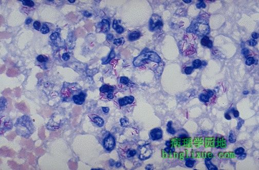 |
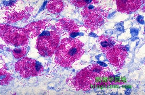
肺结核抗酸染色(MTB)显示结核分枝杆菌,可见红色杆状小体。因此在组织切片或涂片中的结核分枝杆菌(MTB)又称为抗酸杆菌。 This is an acid fast stain of Mycobacterium tuberculosis (MTB). Note the red rods--hence the terminology for MTB in histologic sections or smears: acid fast bacilli. |
 |
分枝杆菌也能被金胺染色,荧光显微镜下抗酸杆菌为发黄*色荧光的杆菌。这个方法易用于筛查分枝杆菌,也常用于实验室痰液标本检查。 Mycobacteria can also be stained with auramine and viewed with fluorescence microscopy, in which acid fast bacilli now appear as glowing yellow rods. This method is easier to use to screen for mycobacteria and is the method routinely used in sputum specimens sent to the laboratory. |
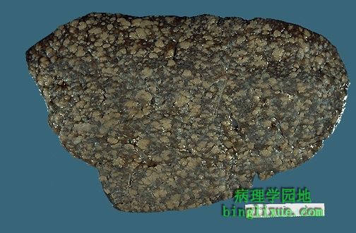 |
脾粟粒性肉芽肿性炎,可见大量黄棕色肉芽肿。意味着免疫反应极差,该病人患AIDS。最后感染被认为是鸟型胞内分枝杆菌( MAI )引起,也即鸟型分枝杆菌复合体( MAC )。 This spleen shows a miliary pattern of granulomatous inflammation, with numerous small tan granulomas. This suggests a poor immune response. This patient had AIDS. The infection turned out to be Mycobacterium avium-intracellulare (MAI), also known as Mycobacterium avium-complex (MAC). |
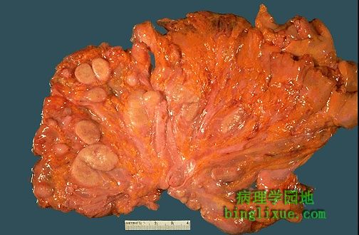 |
左侧见肠系膜上淋巴结,已放大,切面呈黄褐色。淋巴结内充满鸟型分支杆复合体( MAC )。此艾滋病病人免疫反应很差,未形成肉芽肿。 The lymph nodes in this mesentery, best seen at the left, are enlarged and have cut surfaces that appear yellow-tan. These nodes are filled with sheets of Mycobacterium avium-complex (MAC) organisms, and the immune response is so poor in this AIDS patient that there is no focal granuloma formation. |
 |
显微镜下,可见巨噬细胞内大量鸟型分枝杆菌,已用抗酸染色标记出来。抗酸染色后,淋巴结内特别是巨噬细胞内可见大量亮红色杆菌。 Microscopically, Mycobacterium avium-intracellulare infection is marked by numerous acid fast organisms growing within macrophages. Lots of bright red rods are seen, particularly in macrophages, in this acid fast stain of lymph node. |