
 |
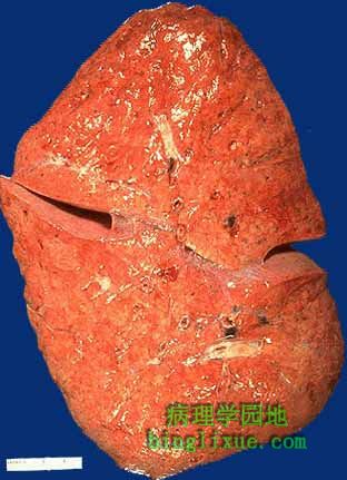
卡氏肺孢子虫肺炎( PCP ),肺出现弥漫性实变,质实如肝。PCP是免疫缺陷病人的典型症状,尤其是AIDS病人。 This lung is as solid as liver because of Pneumocystis carinii pneumonia (PCP). There is diffuse consolidation. PCP is typical of immunocompromised patients, particularly those with AIDS. |
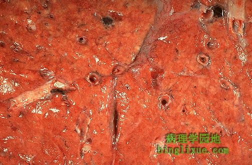 |
卡氏肺孢子虫肺炎( PCP )切面放大后呈橙红色外观。 At higher magnification, the cut surface of the lung with Pneumo- cystis carinii pneumonia (PCP) has a salmon pink appearance. |
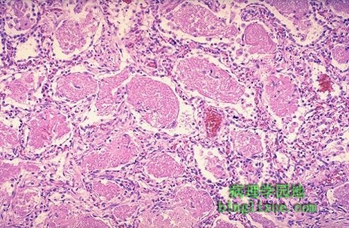 |
显微镜下 ,卡氏肺孢子虫肺炎中每个肺泡内都充满粉红色颗粒状渗出物。 Microscopically, every alveolus is filled with granular pink exudate in this case of Pneumocystis carinii pneumonia. |
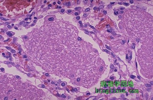 |
高倍镜下,可见卡氏肺孢子虫肺炎肺泡内粉红色颗粒状渗出物。渗出物包含水肿液、蛋白质、肺孢子虫以及坏死的巨噬细胞。这就是气体交换严重障碍的原因。 At higher magnification, the granular pink exudate of Pneumocystis carinii pneumonia is seen. The exudate consists of edema fluid, protein, Pneumocystis organisms, and dead macrophages. One can see why gas exchange is severely compromised. |
 |
诊断卡氏肺孢子虫肺炎最好方法是取肺组织或支气管肺泡灌洗液做GMS染色。孢囊壁被染色后,卡氏肺孢子虫将呈现诸如下列外观,压碎的乒乓球,或新月状,或折叠了的球体,或扁平的沙滩球,或瘪了的网球,等等... The best way to make the diagnosis of Pneumocystis carinii pneumonia is to perform a Gomori methenamine silver (GMS) stain on the lung tissue or bronchoalveolar lavage (BAL) fluid. The cyst wall is stained, and the organisms appear as crushed ping-pong balls, or crescent shapes, or folded spheres, or flattened beach balls, or deflated tennis balls, or.... |
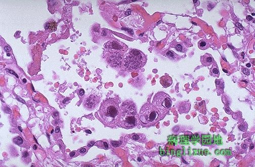 |
巨细胞病毒( CMV )感染的病毒性肺炎,可见体积很大的细胞内有紫色核内包涵体,并且包涵体周围都有一清晰小光晕。胞浆内可见嗜碱性点彩。 This is cytomegalovirus (CMV) infection in the lung. Note the very large cells that have large violet intranuclear inclusions with a small clear halo. Basophilic stippling can be seen in the cytoplasm. |
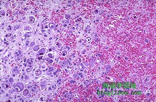 |
肾上腺可见许多巨细胞病毒包涵体,在图片右侧可见坏死和出血。巨细胞病毒通常在免疫缺陷宿主中可见,并在许多器官中蔓延。 Many cytomegalovirus inclusions are seen in this adrenal, which has necrosis and hemorrhage as well at the right. CMV is usually seen in immunocompromised hosts and can be widespread in many organs. |
 |
胎儿先天性巨细胞病毒感染。肾小管上皮内可见巨细胞病毒包涵体。 Here is a congenital cytomegalovirus infection in a fetus. Note the large CMV inclusions in the renal tubular epithelium. |
 |
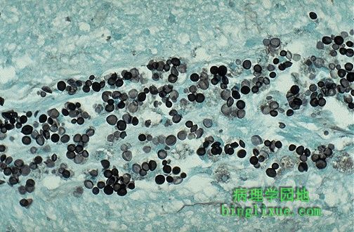
肺新型隐球菌感染,存在许多病原体,每个病原体都有个大粘液囊,此囊在模糊圆形细胞核周围呈现为清晰光亮区。 This is Cryptococcus neoformans infection of the lung. There are numerous organisms that have a large mucoid capsule, giving the appearance of a clear zone around a faint round nucleus. |
 |
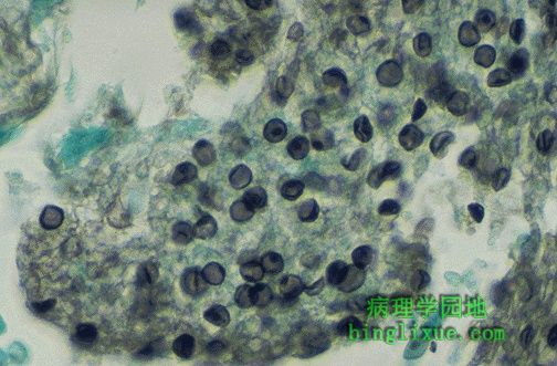
新型隐球菌脑膜炎GMS染色法显示细胞核。爱滋病患者,病原体没形成包囊。隐球菌芽殖细胞的根很细。 This is a Cryptococcus neoformans meningitis stained with GMS to reveal the nuclei. In this AIDS patient, the organisms didn't even bother to make a capsule. The budding cells of Cryptococcus have a narrow base. |