
 |
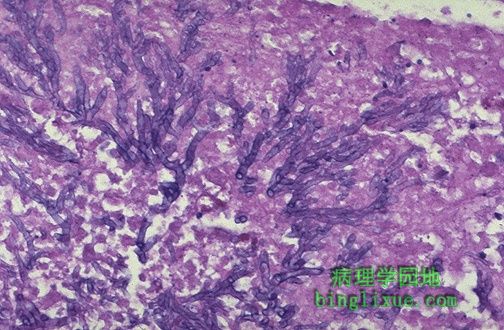
分枝状曲霉菌菌丝。 Here are branching Aspergillus hyphae. |
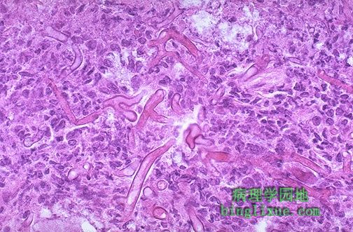 |
宽不分枝的菌丝为毛霉菌的特征。图示部位是糖尿病人的鼻咽部,酮症酸中毒促进真菌的生长。 The broad, non-septate hyphae seen here are characteristic for Mucor. There are several species of zygomycetes, all of which are collectively referred to as the cause for mucormycosis. As in this case, the setting of infection was the nasopharynx in a patient with diabetes mellitus. Ketoacidosis potentiates the growth of this organism. |
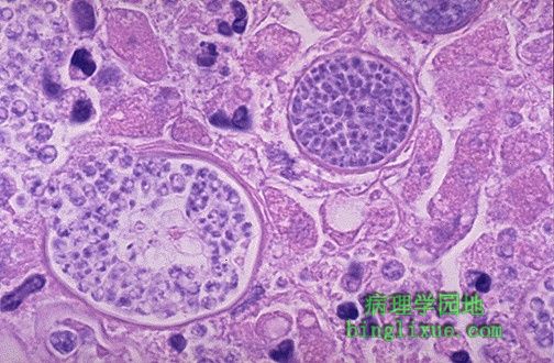 |
充满内生孢子的厚壁大球体是粗球孢子菌感染的特征,是美国西南荒漠特有的。可见左边的大球体破裂并释放出内部的内生孢子,内生孢子生长并引起持续感染。 These large spherules with a thick wall and filled with endospores are characteristic for Coccidioides immitis. "Cocci" is endemic to the desert southwest of the U.S. Note how the spherule at the left is bursting to expel its endospores, which grow and continue the infection. |
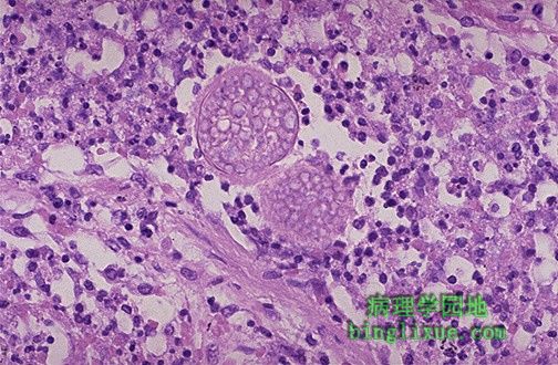 |
粗球孢子菌病肺部感染灶,可见两个大球体充满内生孢子。 This pulmonary infection is due to Coccidioidomycosis, as evidenced by the two large spherules filled with endospores. |
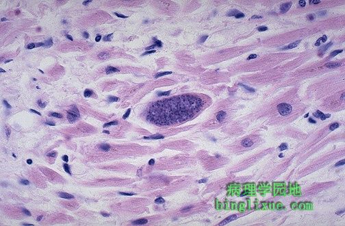 |
爱滋病病人心肌,感染刚地弓形虫假孢囊 。鼠弓形虫感染是免疫缺陷成年人的一种机会性感染,也可能是先天性感染。 This is a Toxoplasma gondii pseudocyst in the myocardium of a patient with AIDS. Toxo is an opportunistic infection of immunocompromised adults. It may also be a congenital infection. |
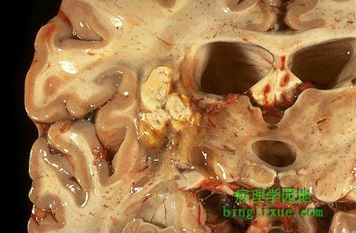 |
脑组织弓形虫脓肿, CT扫描将会呈现为环形病变。 This is a Toxoplasma abscess in the brain, which would appear as a ring-enhancing lesion with CT scan. |
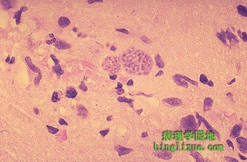 |
艾滋病患者脑组织病理解剖显示小胶质细胞结节内可见刚地弓形虫假孢囊,同时伴有各种炎细胞浸润。 A brain biopsy reveals cysts of Toxoplasma gondii in a microglial nodule with a variety of inflammatory cell types in a patient with AIDS. |
 |
口唇皮肤的清亮小水泡边缘图像。注意存在于表皮上皮细胞的淡紫色及粉红色的核内包涵体。为典型的单纯疱疹病毒( HSV )感染。单纯疱疹病毒感染最常见的部位(原发或继发)是皮肤和粘膜。 HSV I 最常见于口腔, HSV II 主要为性传播疾病。 This is a microscopic section from the edge of one of a group of small round clear vesicles on the skin, just above the lip. Notice the mauve to pink homogenous intranuclear inclusions in the epithelial cells of the epidermis. This is typical for Herpes simplex virus (HSV) infection. The most common sites for Herpes simplex virus infections (either primary or reactivation) are skin and mucus membranes. HSV type I is seen most often in oral cavity, while HSV type II is more commonly a sexually transmitted disease. |
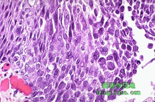 |
食管鳞状上皮可见由单纯疱疹病毒感染引起的境界清楚的溃疡,注意呈紫红色到粉红色的核内包涵体。通常可见多核细胞。此类感染在免疫缺陷病人中最常见。 The esophageal squamous epithelium here is from a sharply demarcated "punched out" ulcer from Herpes simplex virus infection. Note the mauve to pink intranuclear inclusions. Often, multinucleated cells are seen. Such infections are most common in immunocompromised hosts. |
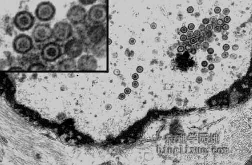 |
单纯疱疹病毒性脑炎,电子显微镜显示大脑神经元内的疱疹病毒颗粒成组排列或单个散在分布。疱疹病毒是外被包膜,核衣壳内含的双股DNA。 By electron microscopy, viral particles of any herpesvirus appear as arrays and scattered single particles as shown here in a nucleus of a neuron from the cerebrum from a patient with herpes simplex encephalitis. Herpesviruses are large encapsulated viruses that contain double-stranded DNA in the nucleocapsid surrounded by the viral envelope. |