
 |
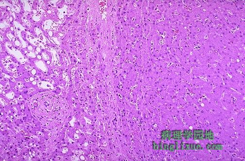
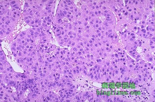
左侧为具有门静脉分支的正常肝组织。右侧为肝脏腺瘤,肿瘤细胞类似于正常肝细胞,但肿瘤组织中肝细胞条索排列紊乱,无正常肝小叶结构。 Normal liver tissue with a portal tract is seen on the left. The hepatic adenoma is on the right and is composed of cells that closely resemble normal hepatocytes, but the neoplastic liver tissue is disorganized hepatocyte cords and does not contain a normal lobular architecture. |
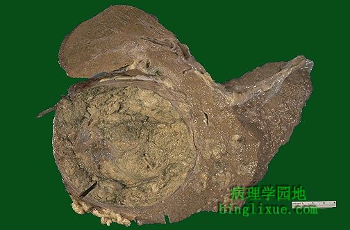 |
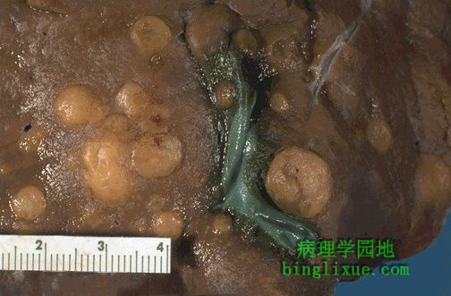
肝细胞癌,可发生于肝硬化。病毒性肝炎是全球最常见的原因,但在美国,最常见的原因为慢性酒精中毒。肿瘤体积巨大,且因含有胆汁而呈绿色。瘤体的右方为小的卫星结节。 Here is an hepatocellular carcinoma. Such liver cancers arise in the setting of cirrhosis. Worldwide, viral hepatitis is the most common cause, but in the U.S., chronic alcoholism is the most common cause. The neoplasm is large and bulky and has a greenish cast because it contains bile. To the right of the main mass are smaller satellite nodules. |
 |
肝细胞癌的卫星结节为肿瘤的肝内扩散或肿瘤的多中心起源。 The satellite nodules of this hepatocellular carcinoma represent either intrahepatic spread of the tumor or multicentric origin of the tumor. |
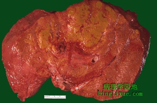 |
呈黄绿色的肝细胞癌。发生肝细胞癌的表现之一为血清甲胎蛋白升高。肿物也可阻塞胆管,导致碱性磷酸酶升高。 Here is another hepatocellular carcinoma with a greenish yellow hue. One clue to the presence of such a neoplasm is an elevated serum alpha-fetoprotein. Such masses may also focally obstruct the biliary tract and lead to an elevated alkaline phosphatase. |
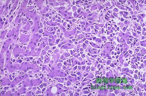 |
该肝细胞癌的恶性肿瘤细胞(主要位于右方)分化较好,并与较大的正常肝细胞(主要位于左方)相互交错。 The malignant cells of this hepatocellular carcinoma (seen mostly on the right) are well differentiated and interdigitate with normal, larger hepatocytes (seen mostly at the left). |
 |
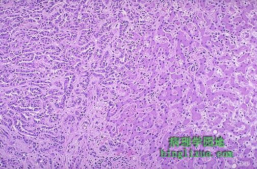
注意此肝细胞癌由宽于正常肝板(厚度为2个细胞)的肝细胞条索组成。虽然血管结构存在,但无可辨认的正常肝小叶结构。 Note that this hepatocellular carcinoma is composed of liver cords that are much wider than the normal liver plate that is two cells thick. There is no discernable normal lobular architecture, though vascular structures are present. |
 |
左侧的肿瘤具有与胆管癌基本相同的腺体表现。肝癌可具有肝细胞分化和胆管分化。胆管癌不产生胆汁,但这些细胞生成粘蛋白,并很难与活检或针吸标本的转移性腺癌相区分。 The carcinoma at the left has a glandular appearance that is most consistent with a cholangiocarcinoma. A liver cancer may have both hepatocellular as well as cholangiolar differentiation. Cholangiocarcinomas do not make bile, but the cells do make mucin, and they can be almost impossible to distinguish from metastatic adenocarcinoma on biopsy or fine needle aspirate. |
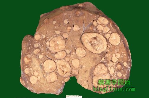 |
大小不等的多个肿块,一些大的肿块中心坏死。很明显这是肝脏转移瘤。肿瘤的阻塞使使碱性磷酸酶水平升高,但它们并未阻塞所有的胆道,因此高胆红素血症不明显。同样,一般没有转氨酶的明显升高。 Note the numerous mass lesions that are of variable size. Some of the larger ones demonstrate central necrosis. The masses are metastases to the liver. The obstruction from such masses generally elevates alkaline phosphatase, but not all bile ducts are obstructed, so hyperbilirubinemia is typically not present. Also, the transaminases are usually not greatly elevated. |
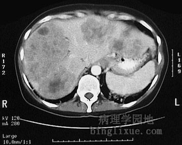 |
腹部横断面CT显示多个肿块使肝脏明显肿大,从左而延伸到右侧。为结肠腺癌肝转移性病变。左下(图右下)可见正常脾。 This computed tomographic (CT) scan without contrast of the abdomen in transverse view demonstrates multiple mass lesions resulting in a markedly enlarged liver extending from right to nearly the left side of the upper abdomen. These are metastases from a colonic adenocarcinoma. A normal sized spleen is seen at the lower left. |
 |
结肠腺癌的肝脏转移癌。结肠是肝脏转移性腺癌的最主要原发部位。 Here are liver metastases from an adenocarcinoma primary in the colon, one of the most common primary sites for metastatic adenocarcinoma to the liver. |