
 |
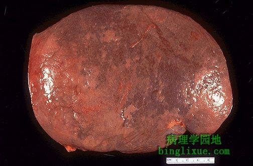
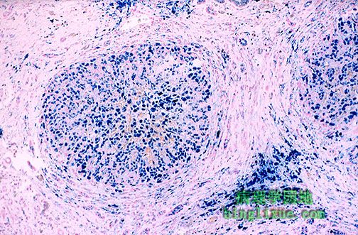
图示,门脉高压的最常见并发症之一脾肿大。脾脏由正常的300克以内增大至500到1000克。此图的另一表现是紫色被膜上遍及胶原的异常淡褐色斑块,称为“糖衣”或“透明蛋白性脾周炎”,它继发于脾脏肿大和/或肝硬化常见并发症腹膜炎反复发作。 One of the most common findings with portal hypertension is splenomegaly, as seen here. The spleen is enlarged from the normal 300 grams or less to between 500 and 1000 gm. Another finding here is the irregular pale tan plaques of collagen over the purple capsule known as "sugar icing" or "hyaline perisplenitis" which follows the splenomegaly and/or multiple episodes of peritonitis that are a common accompaniment to cirrhosis of the liver. |
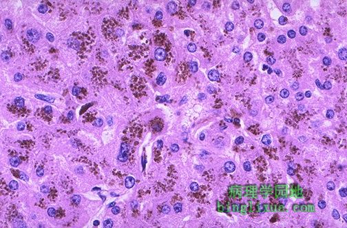 |
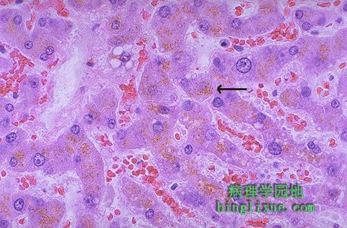
图中肝细胞和枯否细胞内充满由肝脏中过多的铁积聚造成的含铁血黄素的褐色粒状沉着。含铁血黄素沉着表示铁的相对良性积聚。当发生器官功能障碍时,则用“血色素沉着症”表示。铁积聚能够导致小结节性肝硬化(也称为“色素性”肝硬化)。 The hepatocytes and Kupffer cells here are full of granular brown deposits of hemosiderin from accumulation of excess iron in the liver. The term "hemosiderosis" is used to denote a relatively benign accumulation of iron. The term "hemochromatosis" is used when organ dysfunction occurs. The iron accumulation may lead to a micronodular cirrhosis (so called "pigment" cirrhosis). |
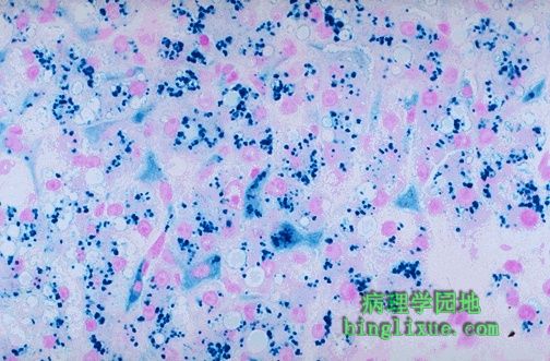 |
普鲁士蓝铁染色显示肝细胞和枯否细胞中含铁血黄素的蓝色颗粒。血色素沉着症或可以是原发的(病因大概为常染色体隐性遗传病),也可以是继发的(过多的铁摄取或吸收、肝脏疾病、大量输血)。血色素沉着症导致皮肤的青铜色色素沉着、糖尿病(累及胰腺)、心率失常(累及心肌)。 A Prussian blue iron stain demonstrates the blue granules of hemosiderin in hepatocytes and Kupffer cells. Hemochromatosis can be primary (the cause is probably an autosomal recessive genetic disease) or secondary (excess iron intake or absorption, liver disease, or numerous transfusions). Hemochromatosis leads to bronze pigmentation of skin, diabetes mellitus (from pancreatic involvement), and cardiac arrhythmias (from myocardial involvement). |
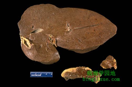 |
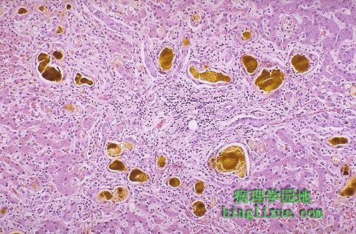
一例遗传性血色素沉着症(HHC),中年男性患者体内发生广泛的铁沉积,导致其肝脏、胰腺(下中图)和淋巴结(下右图)的切面均呈暗褐色。该病是由于血色素沉着症基因(HFE)突变使得肠吸收铁增加所致。美国约1/200-1/500病人发生。1/10的北欧血统的人携带正常的HFE隐性基因,而发病主要是C282Y突变。 The dark brown color of the liver, as well as the pancreas (bottom center) and lymph nodes (bottom right) on sectioning is due to extensive iron deposition in a middle-aged man with hereditary hemochromatosis (HHC). HHC results from a mutation involving the hemochromatosis gene (HFE) that leads to increased iron absorption from the gut. The prevalence is between 1:200 and 1:500 persons in the U.S. About 1 in 10 persons of northern European ancestry carries the abnormal recessive HFE gene, and most of these are the C282Y mutation. |
 |
普鲁士蓝铁染色显示,遗传性血色素沉着症(HH)患者在镜下可见广泛的肝脏含铁血黄素沉着。注意还有肝硬化表现。HH患者过多的铁沉积能够影响许多器官,如心脏(充血性心衰)、胰腺(糖尿病)、肝脏(肝硬化和肝衰)、关节(关节炎)是受累最严重的器官。 The Prussian blue iron stain reveals extensive hepatic hemosiderin deposition microscopically in this case of hereditary hemochromatosis (HH). Note that there is also cirrhosis. Excessive iron deposition in persons with HH can affect many organs, but heart (congestive failure), pancreas (diabetes mellitus), liver (cirrhosis and hepatic failure), and joints (arthritis) are the most severely affected. |
 |
如图所示,存在于几乎全部肝细胞中的淡褐色细小颗粒状色素称为脂色素(脂褐素)。箭头所示为肝细胞的脂褐素沉着。自身吞噬溶酶体长时间积聚形成的“损耗性”色素。该色素不具有实际的病理意义。 The pale golden brown finely granular pigment seen here in nearly all hepatocytes is lipchrome (lipofuscin). One such deposit within a hepatocyte is marked by the arrow. This is a "wear and tear" pigment from the accumulation of autophagolysosomes over time. This pigment is of no real pathologic importance. |
 |
图中微黄绿色的色素积聚为胆汁。这通常由肝外胆道阻塞所引起。但肝细胞损伤时,胆汁在肝脏中积聚(称为胆汁淤积)。 The yellowish-green accumulations of pigment seen here are bile. Most often this is due to extrahepatic biliary tract obstruction. However, bile may also accumulate in liver (called cholestasis) when there is hepatocyte injury. |
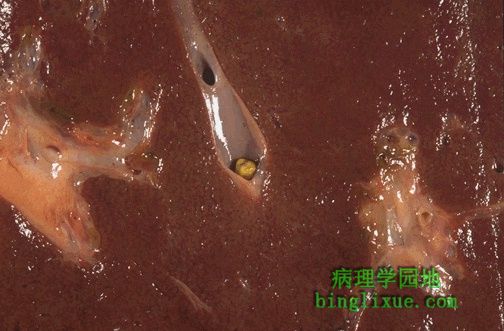 |
肝内胆管小结石引起的肝内阻塞,能够引起局部胆汁淤积,但由于大量未阻塞部分可清除血中胆红素,血清胆红素水平不升高。但任何水平的胆道阻塞时,血清碱性磷酸酶都升高。 Here is an example of intrahepatic obstruction with a small stone in an intrahepatic bile duct. This could produce a localized cholestasis, but the serum bilirubin would not be increased, because there is plenty of non-obstructed liver to clear the bilirubin from the blood. However, the serum alkaline phosphatase is increased with biliary tract obstruction at any level. |
 |
右上方为源于肝脏的境界分明的肿瘤。是肝腺瘤。 At the upper right is a well-circumscribed neoplasm that is arising in liver. This is an hepatic adenoma. |
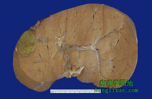 |
肝脏的切面显示了肝脏腺瘤。注意分界清楚。由于慢性酒精中毒引起脂肪变,其余的肝脏呈淡黄褐色。 The cut surface of the liver reveals the hepatic adenoma. Note how well circumscribed it is. The remaining liver is a pale yellow brown because of fatty change from chronic alcoholism. |