
 |
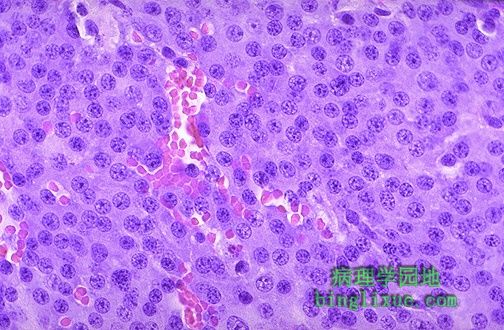
此处显示垂体腺瘤的显微镜图像。可见肿瘤细胞圆而小,形态一致。 The microscopic appearance of the pituitary adenoma is shown here. Note the monotonous appearance of these small round cells. |
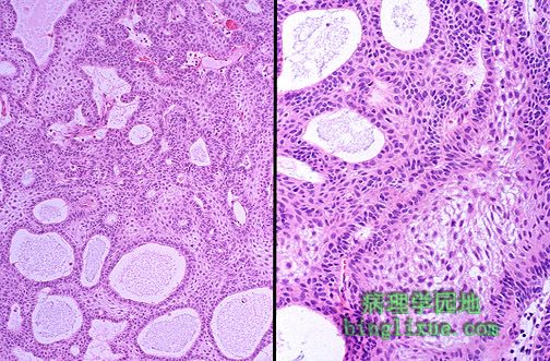 |
中倍镜和高倍镜示颅咽管瘤,起源于Rathke'S囊并且在蝶鞍处形成膨胀性的大肿块,侵蚀骨并浸润周围的组织结构。尽管在组织学上由鳞状上皮和柱状上皮构成囊腔,囊内充满脂性液体,但难以根除。 A craniopharyngioma is seen here at medium and high power. It is derived from remnants of Rathke's pouch and forms an expanding mass arising in the sella turcica that erodes bone and infiltrates into surrounding structures. They are difficult to eradicate, even though they are composed of histologically appearing squamoid and columnar epithelium lining cystic spaces filled with oily fluid. |
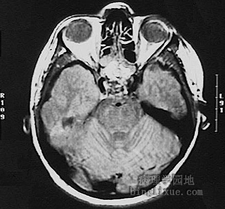 |
头颅CT显示颅咽管瘤,在蝶鞍可见边缘不规则的大肿块,象垂体腺瘤一样压迫视交叉。颅咽管瘤临床少见,多见于青年人。 This head CT scan demonstrates a mass with irregular margins in the region of the sella. Such a mass may impinge upon the optic chiasm, just like a pituitary adenoma. Craniopharyngiomas are uncommon and tend to occur in young persons. |
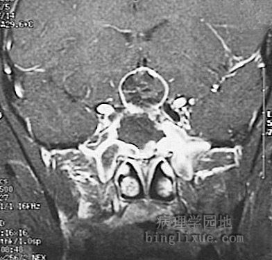 |
颅咽管瘤CT图像(中央靠上边缘为白色的圆形区域) |
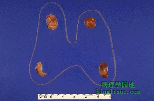 |
图示增生的甲状旁腺,3个半腺体被切除(左下显示半个腺体)。甲状旁腺增生是原发性甲状旁腺功能亢进症第二常见的症状,而甲状旁腺癌最少见。 Parathyroid hyperplasia is shown here. Three and one-half glands have been removed (only half the gland at the lower left is present). Parathyroid hyperplasia is the second most common form of primary hyperparathyroidism, with parathyroid carcinoma the least common form. |
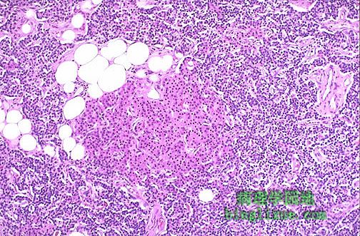 |
甲状旁腺增生很少有或没有脂肪组织存在,但各类细胞正常。可见粉红色的甲状旁腺嗜酸性细胞。实际上是伴有腺体扩大的继发性甲状旁腺功能亢进,它是由慢性肾衰血磷排泄障碍所致。血磷升高使血清钙下降,从而引起甲状旁腺分泌更多的甲状旁腺激素。 In parathyroid hyperplasia, there is little or no adipose tissue, but any or all cell types normally found in parathyroid are present. Note the pink oxyphil cells here. This is actually "secondary hyperparathyroidism" with enlarged glands as a consequence of chronic renal failure with impaired phosphate excretion. The increased serum phosphate tends to drive serum calcium down, which in turn drives the parathyroids to secrete more parathormone. |
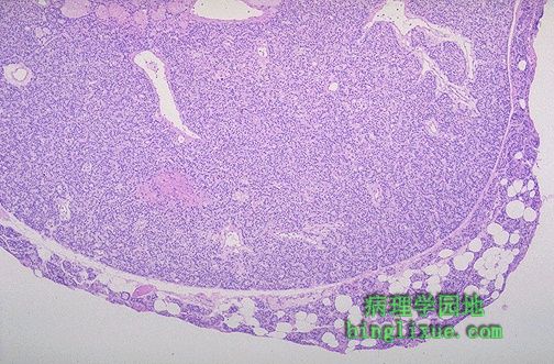 |
甲状旁腺腺瘤是引起原发性甲状旁腺功能亢进最常见的原因。可见混有脂肪细胞的正常甲状旁腺组织的边缘被推向右边及腺瘤下缘。 Here is a parathyroid adenoma, which is the most common cause for primary hyperparathyroidism. A rim of normal parathyroid tissue admixed with adipose tissue cells is seen compressed to the right and lower edge of the adenoma. |
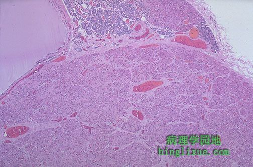 |
右上邻近甲状旁腺腺瘤的是正常甲状旁腺组织(粉红色甲状旁腺嗜酸性细胞结节)的边缘,在左上是一个小的良性甲状旁腺囊肿(意外发现)。患有原发性甲状旁腺功能亢进症的病人通常随身携带电解质常规检测仪,它能随时监测到血钙的高值。甲状旁腺激素( PTH )测定显示PTH升高。 Adjacent to this parathyroid adenoma is a rim of normal parathyroid tissue (with a pink oxyphil cell nodule) at the upper right, and a small benign parathyroid cyst (an incidental finding) is at the upper left. Patients with this form of primary hyperparathyroidism are usually picked up with routine chemistry panels in which a high serum calcium is noted. A parathormone (PTH) assay reveals a high normal to elevated level of PTH. |
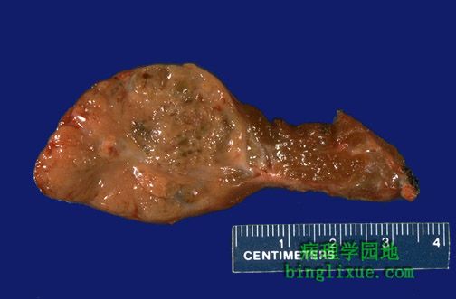 |
甲状旁腺癌的大体外观。血清钙会相当高。可见大而不规则的切面。癌具有侵袭性,可浸润周围组织颈部,使甲状旁腺癌的手术非常复杂。 This is the gross appearance of a parathyroid carcinoma. The serum calcium can be quite high. Note the large size and irregular cut surface. These carcinomas have a tendency to invade surrounding tissues in the neck, complicating their removal. |
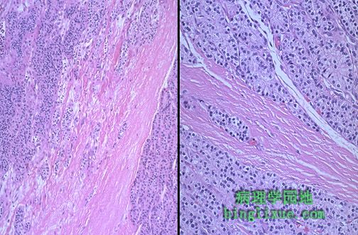 |
左边是甲状旁腺癌中倍镜图像,右边是甲状旁腺癌高倍镜图像。癌巢肿瘤细胞异型性较小。癌巢之间见条索状纤维组织。甲状旁腺癌浸润周围的颈部组织。 This is a parathyroid carcinoma seen at medium power on the left and higher power on the right. The nests of neoplastic cells that are not very pleomorphic. Note the bands of fibrous tissue between the nests. Parathyroid carcinomas infiltrate surrounding structures in the neck. |