
 |
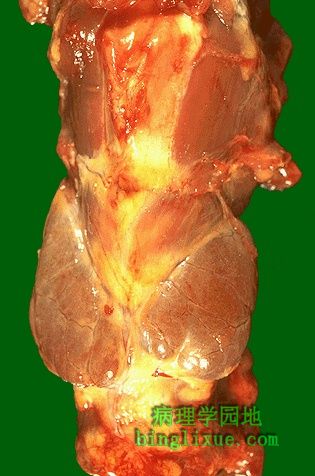
正常甲状腺外观。位于颈部气管前,由左右两叶及中间的峡部构成。正常甲状腺重约10-30g。体检不易摸到。 This is the normal appearance of the thyroid gland on the anterior trachea of the neck. The thyroid gland has a right lobe and a left lobe connected by a narrow isthmus. The normal weight of the thyroid is 10 to 30 grams. It cannot easily be palpated on physical examination. |
 |
镜下,正常甲状腺由滤泡构成。这些滤泡内衬上皮,充满胶质。甲状腺滤泡大小略有差别。间质不明显,其内可见C细胞。 Normal thyroid seen microscopically consists of follicles lined by a an epithelium and filled with colloid. The follicles vary somewhat in size. The interstitium, which may contain "C" cells, is not prominent. |
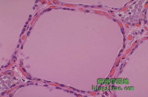 |
正常甲状腺滤泡内衬立方上皮。这些上皮细胞可分泌或重吸收胶质,这有赖于垂体分泌的TSH(促甲状腺激素)的刺激程度。同其他内分泌腺体一样,甲状腺间质富含血管,分泌的激素可进入血管进行运输。 This normal thyroid follicle is lined by a cuboidal follicular epithelium with cells that can add or subtract colloid depending upon the degree of stimulation from TSH (thyroid stimulating hormone) released by the pituitary gland. As in all endocrine glands, the interstitium has a rich vascular supply into which hormone is secreted. |
 |
降钙素抗体免疫过氧化酶染色可见甲状腺C细胞(又称滤泡旁细胞),位于滤泡之间和滤泡上皮细胞之间。C细胞分泌降钙素。 This immunoperoxidase stain with antibody to calcitonin identifies the "C" cells (parafollicular cells) of the thyroid interstitium between the follicles or adjacent to the epithelium of follicles. These cells secrete calcitonin. |
 |
可见对称的萎缩的甲状腺。该病人甲状腺功能低下,此为桥本甲状腺炎的终末病变。最初甲状腺肿大,并出现短暂的甲状腺功能亢进,随后甲状腺功能正常,数年后甲状腺萎缩,出现甲状腺功能减退。异常T细胞激活及随后的B细胞刺激导致自身抗体分泌引起桥本甲状腺炎。 This symmetrically small thyroid gland demonstrates atrophy. This patient was hypothyroid. This is the end result of Hashimoto's thyroiditis. Initially, the thyroid is enlarged and there may be transient hyperthyroidism, followed by a euthyroid state and then hypothyroidism with eventual atrophy years later. Hashimoto's thyroiditis results from abnormal T cell activation and subsequent B cell stimulation to secrete a variety of autoantibodies. |
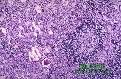 |
低倍镜显示桥本甲状腺炎。右侧中央可见淋巴结。此为自身免疫疾病,通常抗甲状腺球蛋白和微粒体抗体(甲状腺过氧化物酶抗体)能够被检测。其它如Addison病或恶性贫血等自身免疫疾病也可能出现。尽管TSI的抑制性抗体能减轻其效应,仍可见甲状腺生长刺激免疫球蛋白( TGI ) 和甲状腺刺激免疫球蛋白( TSI )。 Here is a low power microscopic view of a thyroid with Hashimoto's thyroiditis. Note the lymphoid follicle at the right center. This is an autoimmune disease and often antithyroglobulin and antimicrosomal (thyroid peroxidase) antibodies can be detected. Other autoimmune diseases such as Addison's disease or pernicious anemia may also be present. Both thyroid growth immunoglobulins (TGI) and thyroid stimulating immunoglobulins (TSI) are present, though blocking antibodies to TSI mitigate their effect. |
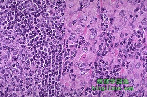 |
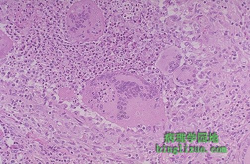
高倍镜下示桥本甲状腺炎。甲状腺中心部位和右边可见粉红色的hürthle细胞。左边是淋巴结。桥本甲状腺炎开始引起无痛性甲状腺肿大,数年后出现甲状腺萎缩。 This high power microscopic view of the thyroid with Hashimoto's thyroiditis demonstrates the pink Hürthle cells at the center and right. The lymphoid follicle is at the left. Hashimoto's thyroiditis initially leads to painless enlargement of the thyroid, followed by atrophy years later. |
 |
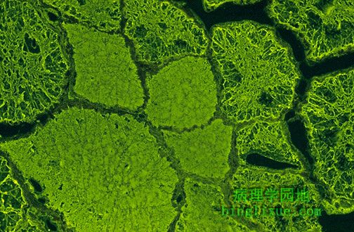
抗微粒体抗体免疫荧光法阳性的病例,为甲状腺自身免疫疾病中自身抗体之一。抗微粒体抗原主要组成之一是甲状腺过氧化物酶( TPO ),它可通过血清学方法检测出来。注意显示为亮绿色荧光的甲状腺上皮细胞,而位于滤泡中心的胶质颜色较暗。 This is an example of an immunofluorescence test positive for anti-microsomal antibody, one of the autoantibodies that can be seen with autoimmune diseases of the thyroid. A major component of the antimicrosomal antigen is thyroid peroxidase (TPO) which is often measured serologically. Note the bright green fluorescence in the thyroid epithelial cells, whereas the colloid in the center of the follicles is dark. |
 |
抗甲状腺球蛋白免疫荧光法阳性的病例。病人患有桥本甲状腺炎,也可能患有其它自身免疫疾病,如 Graves病、系统性红斑狼疮、类风湿性关节炎,恶性贫血症,以及口眼干燥综合症。 Here is an example of immunofluorescence positivity for anti-thyroglobulin antibody. Patients with Hashimoto's thyroiditis may also have other autoimmune conditions including Grave's disease, SLE, rheumatoid arthritis, pernicious anemia, and Sjogren's syndrome. |
 |
亚急性肉芽肿性甲状腺炎(Dequervain病),它的发生可能与病毒感染有关。甲状腺肿大伴有疼痛。为自限性疾病,病程为数周到数月不等,过后甲状腺功能恢复正常。可见破坏甲状腺滤泡的异物巨细胞。 This is subacute granulomatous thyroiditis (DeQuervain's disease), which probably follows a viral infection and leads to a painful enlarged thyroid. This disease is usually self-limited over weeks to months and the patients return to a euthyroid state. Note the foreign body giant cells with destruction of thyroid follicles. |