
 |
甲状腺大小正常,但左下极可见一个大的胶样囊肿,右下极也有一小胶样囊肿。这种囊肿在甲状腺扫描时显示为“冷”结节。它们是较少见的良性病变,形成的肿块可以与可能的甲状腺癌分辨出来。 This thyroid gland is about normal in size, but there is a larger colloid cyst at the left lower pole and a smaller colloid cyst at the right lower pole. Such cysts could appear as "cold" nodules on a thyroid scan. They are incidental benign lesions but can appear as a mass to be distinguished from possible carcinoma. |
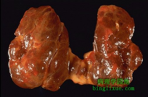 |
弥漫性甲状腺肿在一定程度上是结节状。病人甲状腺功能正常。结节性甲状腺肿是甲状腺肿大最常见的原因,也是最常见的甲状腺疾病。 This diffusely enlarged thyroid gland is somewhat nodular. This patient was euthyroid. This represents the most common cause for an enlarged thyroid gland and the most common disease of the thyroid--a nodular goiter. |
 |
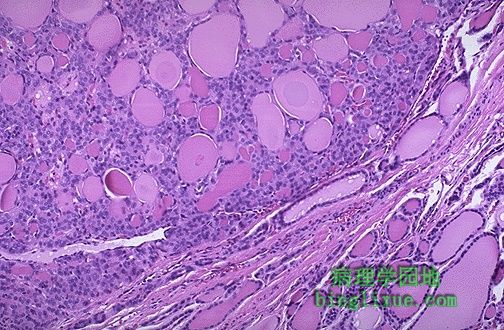
多结节性甲状腺肿低倍镜图像。滤泡不规则增大,扁平上皮组织处于非增殖状态。弥漫性(非毒性)甲状腺肿早期就是这种病理变化,可能的病因如下,地方性甲状腺肿(在一些饮食缺碘地区可能发生)或少见的“非地方性”或散发性甲状腺肿 (年轻的成年女性多发)。甲状腺激素先天性合成异常引起的甲状腺肿极为少见。 The follicles are irregularly enlarged, with flattened epithelium, consistent with inactivity, in this microscopic appearance at low power of a multinodular goiter. The earlier phase of a diffuse (non-toxic) goiter leading up to this point may have resulted from either "endemic" goiter (seen in parts of the world where dietary deficiency of iodine may occur) or the uncommon "nonendemic" or sporadic goiter (young adult women are most often affected). Inborn errors of thyroid hormone biosynthesis leading to goiter are extremely uncommon. |
 |
Graves病典型的病变是甲状腺弥漫性对称性肿大,当然伴有甲状腺功能亢进。低倍镜下,滤泡上皮呈乳头状增生。在这类自身免疫病中,甲状腺刺激免疫球蛋白(TSI)的作用远强于甲状腺生长刺激免疫球蛋白(TGI)的作用。 A diffusely enlarged thyroid gland associated with hyperthyroidism is known as Grave's disease. At low power here, note the prominent infoldings of the hyperplastic epithelium. In this autoimmune disease the action of TSI's predominates over that of TGI's. |
 |
高倍镜下,Graves病时甲状腺增生上皮呈高柱状,内摺排列融进胶质。临近上皮细胞的胶质内可见清晰空泡,此处上皮组织细胞活跃,甲状腺激素分泌增多,形成扇形的胶质边缘。 At high power, the tall columnar thyroid epithelium with Grave's disease lines the hyperplastic infoldings into the colloid. Note the clear vacuoles in the colloid next to the epithelium where the increased activity of the epithelium to produce increased thyroid hormone has led to scalloping out of the colloid. |
 |
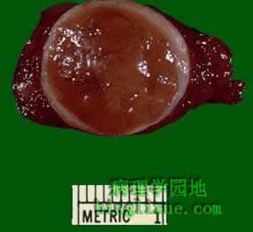
甲状腺手术切除标本,为一小肿块,被切为两半。在左边可见模糊的甲状腺实质边缘。肿块边界清楚。质地非常坚韧。采用]闪烁扫描法扫描呈冷结节。图为滤泡状腺瘤。 Here is a surgical excision of a small mass from the thyroid gland that has been cut in half. A rim of slightly darker thyroid parenchyma is seen at the left. The mass is well-circumscribed. Grossly it felt firm. By scintigraphic scan it was "cold." This is a follicular adenoma. |
 |
另例滤泡状腺瘤,有薄层白色包膜包绕。有时很难区分分化好的滤泡状腺癌与滤泡状腺瘤。因此,对滤泡状肿瘤采取甲状腺次全切是比较安全的措施。 Here is another follicular neoplasm (a follicular adenoma histologically) that is surrounded by a thin white capsule. It is sometimes difficult to tell a well-differentiated follicular carcinoma from a follicular adenoma. Thus, patients with follicular neoplasms are treated with subtotal thyroidectomy just to be on the safe side. |
 |
右下是正常的甲状腺滤泡。左上为滤泡状腺瘤。腺瘤很接近于正常组织结构,属于分化较好的肿瘤。腺瘤滤泡内包含胶质,但滤泡大小不等。 Normal thyroid follicles appear at the lower right. The follicular adenoma is at the center to upper left. This adenoma is a well- differentiated neoplasm because it closely resemble normal tissue. The follicles of the adenoma contain colloid, but there is greater variability in size than normal. |
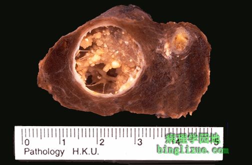 |
甲状腺乳头状癌。在甲状腺内易侵袭淋巴管,因此淋巴结转移较常见,可见多个病灶。肿块呈囊性,内部包含乳头状赘生物。最常见于中年女性。 Sectioning through a lobe of excised thyroid gland reveals papillary carcinoma. This neoplasm can be multifocal, as seen here, because of the propensity to invade lymphatics within thyroid, and lymph node metastases are common. The larger mass is cystic and contains papillary excresences. These tumors most often arise in middle-aged females. |
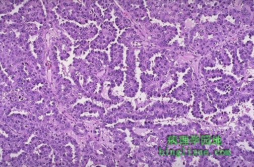 |
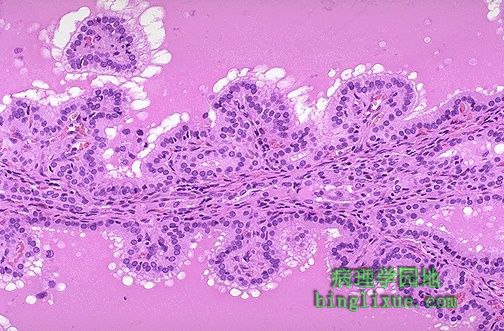
甲状腺乳头状癌组织学外观。乳头分枝多,乳头中心有薄层纤维血管间质。再没有其它肿瘤与乳头状腺瘤结构相同,并且所有的甲状腺乳头状瘤都应以恶性看待。 This is the microscopic appearance of a papillary carcinoma of the thyroid. The fronds of tissue have thin fibrovascular cores. The fronds have an overal papillary pattern. There is no such thing as a papillary adenoma, and all papillary neoplasms of the thyroid should be considered malignant. |