
 |
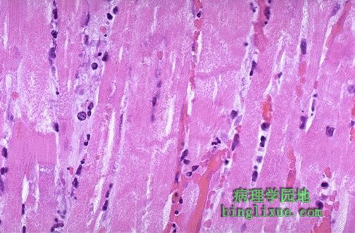
高倍镜示心肌梗死大约1到2天。心肌纤维有暗红的收缩带经过,心肌细胞核几乎全部消失,有急性炎症的迹象,急性心肌梗死在临床上表现为心电图改变以及肌酸激酶同工酶升高。 This high power microscopic view of the myocardium demonstrates an infarction of about 1 to 2 days in duration. The myocardial fibers have dark red contraction bands extending across them. The myocardial cell nuclei have almost all disappeared. There is beginning acute inflammation. Clinically, such an acute myocardial infarction is marked by changes in the electrocardiogram and by a rise in the MB fraction of creatine kinase. |
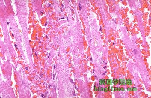 |
心肌梗死不久可见广泛出血合并心肌纤维坏死以及细胞核消失。 In this microscopic view of a recent myocardial infarction, there is extensive hemorrhage along with myocardial fiber necrosis with contraction bands and loss of nuclei. |
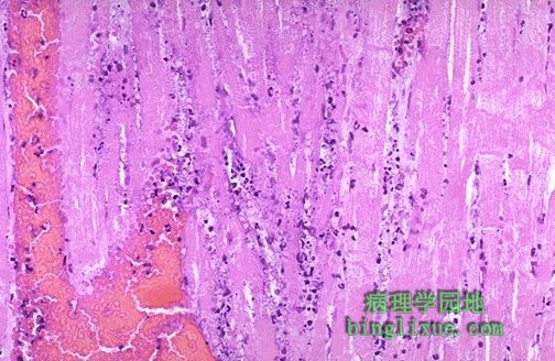 |
心肌梗死大约3到4天可见许多急性炎症细胞浸润,心肌纤维坏死严重仅能见到其模糊轮廓。 This myocardial infarction is about 3 to 4 days old. There is an extensive acute inflammatory cell infiltrate and the myocardial fibers are so necrotic that the outlines of them are only barely visible. |
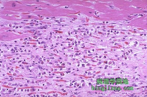 |
中度心肌梗死一到两周,图上方残存一些正常的心肌纤维,在这些纤维下方可见许多巨噬细胞以及丰富的毛细血管和少许胶原形成。 This is an intermediate myocardial infarction of 1 to 2 weeks in age. Note that there are remaining normal myocardial fibers at the top. Below these fibers are many macrophages along with numerous capillaries and little collagenization. |
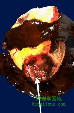 |
透壁性心肌梗塞并发症之一是心脏破裂,极有可能在初次发作后的第三至第五天发生,因此时心肌最柔软。在左心室前壁和室间隔前部,白色箭头所指的即为破裂点。注意暗红血凝块为心包积血,可致心包填塞。 One complication of a transmural myocardial infarction is rupture of the myocardium. This is most likely to occur in the first week between 3 to 5 days following the initial event, when the myocardium is the softest. The white arrow marks the point of rupture in this anterior-inferior myocardial infarction of the left ventricular free wall and septum. Note the dark red blood clot forming the hemopericardium. The hemopericardium can lead to tamponade. |
 |
横切面箭头示心肌的破裂点。三周前发生过一次心肌梗死,相应部位心室壁非常薄弱,因此第二次心肌梗死发生三天后出现破裂。 In cross section, the point of rupture of the myocardium is shown with the arrow. In this case, there was a previous myocardial infarction 3 weeks before, and another myocardial infarction occurred, rupturing through the already thin ventricular wall 3 days later. |
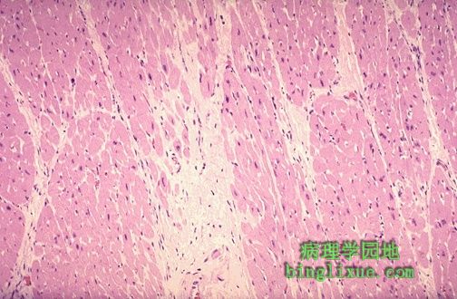 |
心肌纤维之间可见淡白色的胶原,表明为陈旧性梗死特征。 There is pale white collagen within the interstitium between myocardial fibers. This represents an area of remote infarction. |
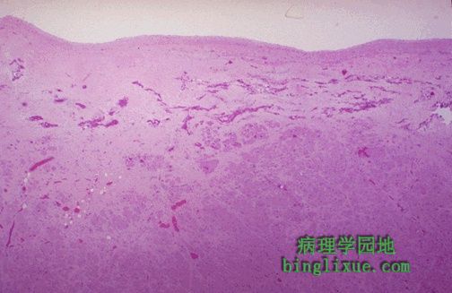 |
图上方心内膜下的心肌可见苍白色纤维化,是心内膜下心肌梗死治疗后形成的。 The myocardium beneath the endocardial surface at the top demonstrates pale fibrosis with collagenization following healing of a subendocardial myocardial infarction. |
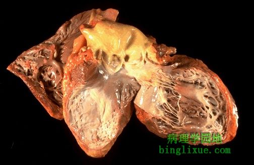 |
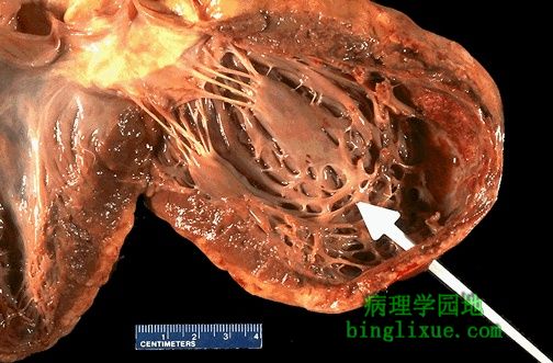
剖开心脏见右侧为左心室壁,中间为室间隔。左心室壁前部以及室间隔广泛地存在陈旧性心肌梗死,心内膜表面的白色区域表示大片瘢痕。 The heart is opened to reveal the left ventricular free wall on the right and the septum in the center. There has been a remote myocardial infarction that extensively involved the anterior left ventricular free wall and septum. The white appearance of the endocardial surface indicates the extensive scarring. |
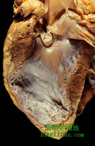 |
左心室壁见大片陈旧性透壁性梗死。尽管心肌壁的厚度正常,但内部仅为薄弱纤维。由于梗死范围太大,以至愈后心室壁被胶原纤维所取代,形成动脉瘤。动脉瘤弹性差,降低了心脏的每搏排出量并增大了残余心肌张力。 There has been a previous extensive transmural myocardial infarction involving the free wall of the left ventricle. Note that the thickness of the myocardial wall is normal superiorly, but inferiorly is only a thin fibrous wall. The infarction was so extensive that, after healing, the ventricular wall was replaced by a thin band of collagen, forming an aneurysm. Such an aneurysm represents non-contractile tissue that reduces stroke volume and strains the remaining myocardium. The stasis of blood in the aneurysm predisposes to mural thrombosis. |