
 |
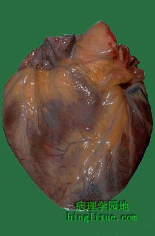
This is the external appearance of a normal heart.The epicardial surface is smooth and glistening.The amount of epicardial fat is usual.The left anterior descending coronary artery extends down from the aortic root to the apex. 图示:正常心脏 心外膜光滑有光泽,存在一些脂肪是正常的。左冠状动脉前降支从主动脉根部发出。 |
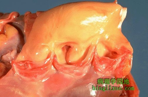 |
The aortic valve shows three thin and delicate cusps. The coronary artery orifices can be seen just above.The endocardium is smooth, beneath which can be seen a red-brown myocardium. The aorta above the valve displays a smooth intima with no atherosclerosis. 主动脉瓣由三个薄而精细的瓣叶组成。在上方的主动脉上可以看到冠状动脉的开口。光滑的心内膜下面是红棕色的心肌。主动脉瓣上方的血管内膜光滑,不伴有动脉粥样硬化。 |
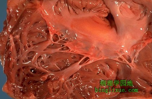 |
This is the tricuspid valve. The leaflets and thin and delicate. Just like the mitral valve, the leaflets have thin chordae tendineae that attach the leaflet margins to the papillary muscles of the ventricular wall below. 图示三尖瓣,瓣叶薄而精巧。瓣叶借腱索与室壁上的乳头肌相连。 |
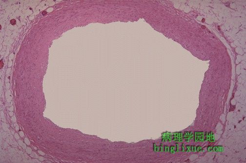 |
This is a normal coronary artery. The lumen is large, without any narrowing by atheromatous plaque. The muscular arterial wall is of normal proportion. 图示:正常冠状动脉。腔较大,不伴有动脉粥样硬化斑块引起的管腔狭窄。动脉肌层的厚度适中。 |
 |
This is the normal appearance of myocardial fibers in longitudinal section. Note the central nuclei and the syncytial arrangement of the fibers, some of which have pale pink intercalated disks. 图示:正常心肌纵切面 可以看到细胞核和平等排列的心肌纤维,心肌纤维间的线形粉红色条纹为闰盘。 |
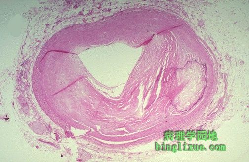 |
The coronary artery shown here has narrowing of the lumen due to build up of atherosclerotic plaque. Severe narrowing can lead to angina, ischemia, and infarction. 冠状动脉粥样梗化使管腔狭窄,严重狭窄时由于缺血可致心绞痛甚至心肌梗死。 |
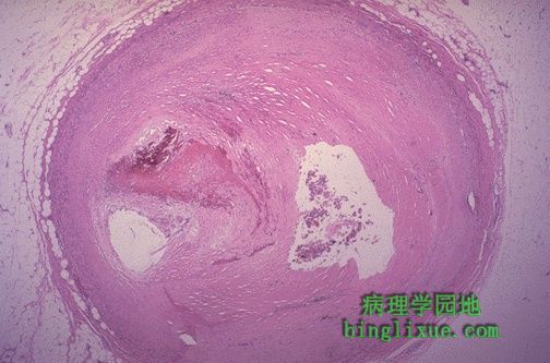 |
This section of coronary artery demonstrates remote thrombosis with recanalization to leave only two small, narrow channels. 图示:冠状动脉血栓形成后再通。可以看到两个狭窄的通道。 |
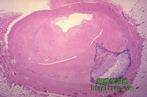 |
There is a severe degree of narrowing in this coronary artery. It is "complex" in that there is a large area of calcification on the lower right, which appears bluish on this H&E stain. Complex atheroma have calcification, thrombosis, or hemorrhage. Such calcification would make coronary angioplasty difficult. 图示:严重的冠状动脉粥样硬化。右下方的HE染色呈蓝色污斑的即为钙化灶。常见的复合病变有钙化、血栓形成、出血等。象这种严重的钙化将使冠脉扩张(手术)带来困难。 |
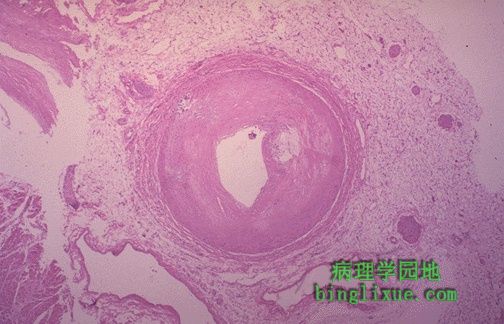 |
This distal portion of coronary artery shows significant narrowing. Such distal involvement is typical of severe coronary atherosclerosis, such as can appear with diabetes mellitus or familial hypercholesterolemia. This would make a coronary bypass operation difficult. 冠状动脉的小分支明显狭窄,是典型的严重的冠状动脉粥样硬化(如可出现于糖尿病和家族性高脂血症患者)的结果。这将为冠状动脉搭桥太造成困难。 |
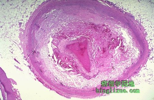 |
There is a pink to red recent thrombosis in this narrowed coronary artery. The open, needle-like spaces in the atheromatous plaque are cholesterol clefts. 在狭窄的冠状动脉中新近形成了血栓。在粥样斑块中看到的针状空隙即为胆固醇结晶。 |