
 |
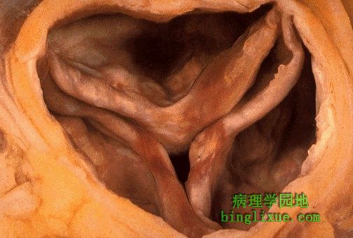
主动脉瓣并非是两个瓣叶的才钙化。有时,在老年人中正常的三个瓣叶的主动脉瓣也会发生钙化,被称为“老年性钙化的主动脉瓣狭窄”。可见瓣膜尖端有钙化结节。 An aortic valve need not be bicuspid to calcify. Sometimes in older adults, a normal tricuspid aortic valve will undergo calcification, a so-called "senile calcific aortic stenosis." Nodules of calcification are seen on the cusps here. |
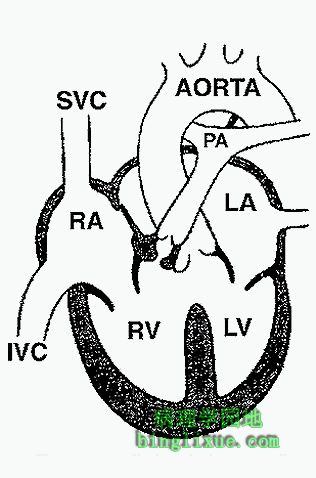 |
图示法乐氏四联征:1.室间隔缺损;2.主动脉骑跨;3.肺动脉狭窄;4.右心室肥大。右心室肥大引起右向左分流,导致发绀。 This diagram depicts the features of Tetralogy of Fallot:1. Ventricular septal defect; 2. Overriding aorta; 3. Pulmonic stenosis; 4. Right ventricular hypertrophy. The obstruction to right ventricular outflow creates a right-to-left shunt that leads to cyanosis. |
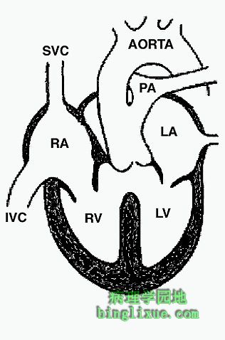 |
图示永存动脉干,见于闭合不全以及动脉干和动脉圆锥(正常情况下将主动脉和肺动脉分开)的旋脊下降不够。一旦螺旋形隔板下降不全,主动脉和肺动脉干的血流将不能分开。这造成左右心室的动脉骑跨。永存动脉干常常伴发膜性室间隔缺损。 The diagram above depicts the findings with a persistent truncus arteriosus. This occurs when there is failure of fusion and descent of the spiral ridges of the truncus and conus that would ordinarily divide into aorta and pulmonic trunck respectively. When the spiral septum fails to completely descend, the aortic and pulmonic trunks are left undivided at their outflow.The truncus overrides both ventricles.The persistent truncus is always accompanied by a membranous ventricular septal defect. |
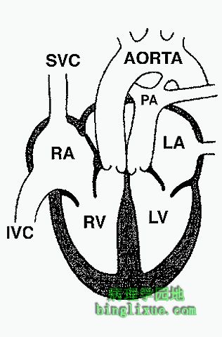 |
图示主动脉肺动脉错位。它在动脉干隔膜非螺旋而是呈直线下降时发生。结果导致右心室的血流进入主动脉而左心室的血流则进入肺动脉。为了维持整个循环系统的正常运行,在休循环和肺循环之间一定存在联系。有时通过室间隔缺损或房间隔缺损相通。在左图,则通过未闭的动脉导管相通。 In the diagram above, transposition of the great vessels is shown. This occurs when the trunco-conal septum does not spiral down. Instead, it descends straight down. As a result, the outflow of right ventricle is into the aorta and the outflow from the left ventricle is into the pulmonic trunk.In order for this system to work, there must be a connection between the system and pulmonic circulations. Sometimes this is through a ventricular septal defect or an atrial septal defect. In the diagram at the left, this is through a patent ductus arteriosus. |
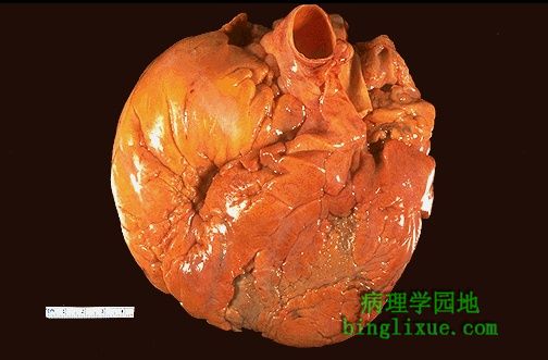 |
球形心,全部房室都扩张。摸上去非常软,心肌收缩力下降。这是心肌病的例子。本例心肌功能下降以及心脏变大和扩张,但未见特征性的组织学改变。 This very large heart has a globoid shape because all of the chambers are dilated. It felt very flabby, and the myocardium was poorly contractile. This is an example of a cardiomyopathy. This term is used to denote conditions in which the myocardium functions poorly and the heart is large and dilated, but there is no specific histologic finding. |
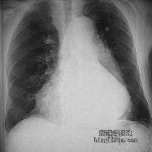 |
胸片示心脏肥大。 This chest radiograph demontrates marked cardiomegaly, with the left heart edge appearing far to the left. |
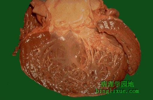 |
这是一个巨大、扩张的左心室,它是扩张性(也叫充血性)心肌病的典型特征。很多病例无明显的病因(因此被称为“原发性扩张性心肌病”),其它的病例则可能与长期酗酒有关。整个心脏变得又大又软。 Here is a large, dilated left ventricle typical of a dilated, or congestive, cardiomyopathy. Many of these have no known etiology (so-called "idiopathic dilated cardiomyopathy") while others may be associated with chronic alcoholism. The heart is very enlarged and flabby. |
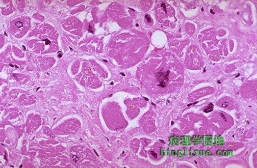 |
镜下可见有心肌病心脏的心肌纤维(同时可见明显的染色较深的细胞核)肥大,还伴有心肌间质纤维化。 Microscopically, the heart in cardiomyopathy demonstrates hypertrophy of myocardial fibers (which also have prominent dark nuclei) along with interstitial fibrosis. |
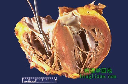 |
左心室明显肥厚,同时可见巨大室间隔有非对称隆起伸入左心室腔内。图为肥厚性心肌病。虽然有大约一半的病例是家族性的,但是引起本病可能是多个基因改变所致。儿童和成年人都可发病,也可引发猝死。请看这个移植心脏,起搏器金属丝深入到右心室。与静脉及大血管相连的心房从后联接到移植心(心脏的供者经过充分的护理,以使得移植有可能成功)。 There is marked left ventricular hypertrophy, with asymmetric bulging of a very large interventricular septum into the left ventricular chamber. This is hypertrophic cardiomyopathy. About half of these cases are familial, though a variety of different genes may be responsible for this disease. Both children and adults can be affected, and sudden death can occur. Seen here is the explanted heart. Pacemaker wires enter the right ventricle. The atria with venous connections, along with great vessels, remained behind to connect to the transplanted heart (provided by someone who cared enough to make transplantation possible). |
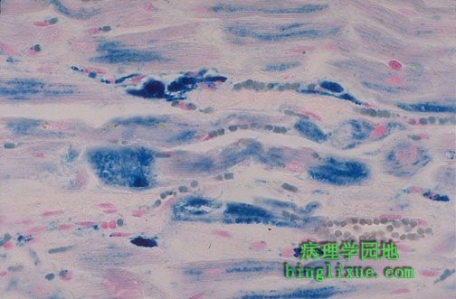 |
血色素沉着症是由于铁过度的沉积而引起的。如图所示,经过普鲁士蓝铁染色后可见该病显微镜下的表现。铁的过度沉淀可导致心脏增大以及类似心肌病的心力衰竭,这使得血色素沉着症成为限制性心肌病的一种形式。 Hemochromatosis, with excessive iron deposition, can occur in the heart as shown here microscopically with Prussian blue iron stain. The excessive deposition of iron leads to heart enlargement and failure similar to a cardiomyopathy, making hemochromatosis a form of "restrictive" cardiomyopathy. |