
 |
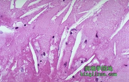
The cholesterol clefts of lipid, along with a few scattered foam cells and a couple of lymphocytes, are seen at high magnification in this atheromatous plaque. 高倍镜下粥样斑块中可见胆固醇结晶空隙,周围有一些散在的泡沫细胞和少量的淋巴细胞 |
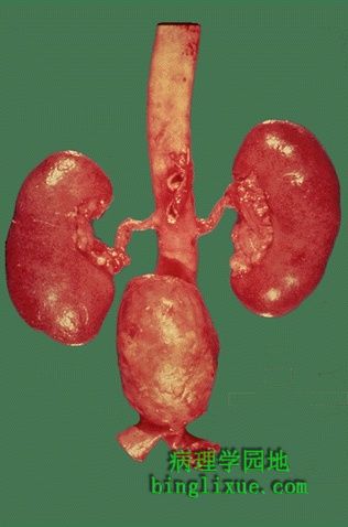 |
Atherosclerosis may weaken the wall of the aorta such that it bulges out to form an aneurysm. An atherosclerotic aortic aneurysm typically occurs in the abdominal portion below the renal arteries, as shown here. Aortic aneurysms that get bigger than 6 or 7 cm are likely to rupture. 动脉粥样硬化处的动脉壁变得很薄弱,容易膨出形成动脉廇。图示:肾动脉以下的腹主动脉廇。大动脉廇的直径大于6到7cm时很容易引起破裂。 |
 |
This is a different kind of arteriosclerosis. This is hyperplastic arteriolosclerosis, which most often appears in the kidney in patients with malignant hypertension. The arteriolar wall is markedly thickened and the lumen is narrowed. 这是另一种类型的动脉硬化的表现,呈增生性细动脉硬化,常见于恶性高血压引起的肾动脉硬化。动脉壁明显增厚,管腔狭窄。 |
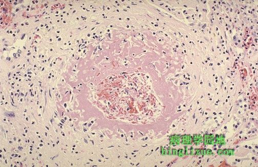 |
Sometimes the small arteries and arterioles can be damaged so severely in malignant hypertension that they demonstrate necrosis with a pink fibrin-like quality that gives this process its name--fibrinoid necrosis. 有时恶性高血压小动脉和细动脉的病变也是非常严重的。呈现伴有粉红色的纤维素样物质形成的坏死(纤维素样坏死)。 |
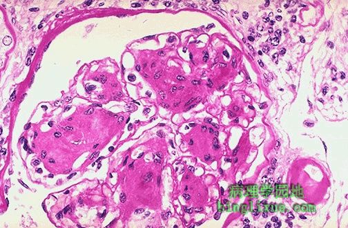 |
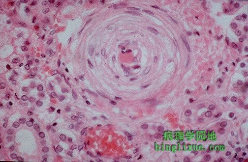
In diabetics, hyaline arteriolosclerosis is common. The glomerulus here stained with PAS shows nodular deposits of amorphous material (nodular glomerulosclerosis) along with a thickened arteriole at the lower right. 糖尿病患者,细动脉玻璃样硬化(变性)是很常见的。这个PAS染色的肾小球可见右下角处的增厚的细动脉管壁有无定形的物质沉积。 |
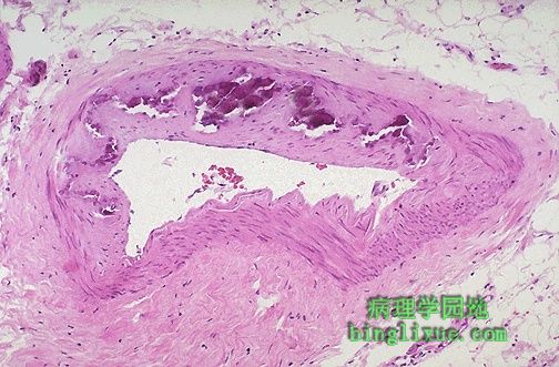 |
This is Monckeberg's medial calcific sclerosis, which is the most insignificant form of arteriosclerosis (both atherosclerosis and arteriolosclerosis are definitely significant). Note the purplish blue calcifications in the media; note that the lumen is unaffected by this process. Thus, there are no real clinical consequences. Remember this process when calcified muscular arteries show up on a radiograph of the pelvic region in an older person. Monckeberg氏动脉中层钙化硬化(动脉中层钙化)是另一种不重要的硬化形式(动脉粥样硬化和细动脉硬化是两种重要的形式)。在动脉中膜可见紫蓝色的钙化灶,而管腔没有受累。看不到明显临床表现。在老年人的X线上骨盆区可以见到肌层钙化的动脉。 |
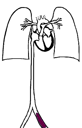 |
A pulmonary thromboembolus travels from a large vein in the leg up the inferior vena cava to the main pulmonary arteries as they branch. Such thrombi embolize most often from large veins in the legs and pelvis where thrombi form with stasis. 肺血栓栓子的运行途径:从下肢静脉向上到达肺动脉主干及其分支,血栓栓子常来自下肢和盆腔的大静脉。 |
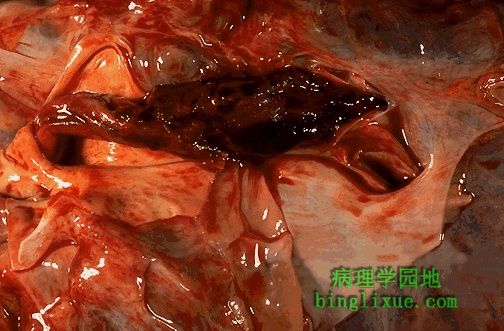 |
The main pulmonary trunk and pulmonary arteries to right and left lungs are seen here opened to reveal a large "saddle" pulmonary thromboembolus. Such an embolus will kill your patient. 可见剖开的肺动脉主干和左肺动脉右肺动脉内有一个大的鞍状的肺血栓栓子。这种栓子可导致病人死亡。 |
 |
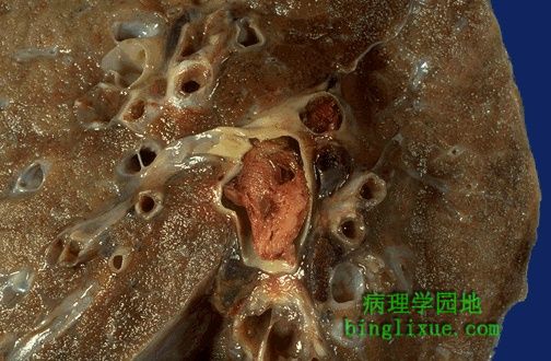
Here is another large pulmonary thromboembolus seen in cross section of this lung. The typical source for such thromboemboli is from large veins in the legs and pelvis. 切面上可见一个大的肺血栓栓子。它的主要来源是下肢和盆腔的大静脉。 |
 |
This pulmonary thromboembolus is occluding the main pulmonary artery. Persons who are immobilized for weeks are at greatest risk. The patient can experience sudden onset of shortness of breath. Death may occur within minutes. 这个肺血栓栓子使肺动脉主干闭塞。数周不动的人很有可能发生。病人可突然出现呼吸急促。数分钟内可引起死亡。 |