
 |
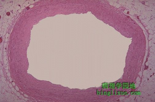
This is a normal coronary artery with no atherosclerosis and a widely patent lumen that can carry as much blood as the myocardium requires. 图示:正常冠状动脉。 无粥样硬化,内腔较大,供血可完全满足心肌的需要。 |
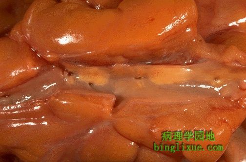 |
This is mild coronary atherosclerosis. A few scattered yellow lipid plaques are seen on the intimal surface of the opened coronary artery traversing the epicardial surface of a heart. The degree of atherosclerosis here is not significant enough to cause disease, but could be the harbinger of worse atherosclerosis to come. 图示:轻度动脉粥样硬化。 在冠状动脉内膜表面可见散在的黄*色脂质斑块。这种程度的动脉粥样硬化不会引起明显的疾病,但它是动脉粥样硬化恶化的先兆。 |
 |
The degree of atherosclerosis is much greater in this coronary artery, and the lumen is narrowed by half. A small area of calcification is seen in the plaque at the right. 图示:较重的冠状动脉粥样硬化。 内腔仅有正常的一半,右边可见钙化斑块。
|
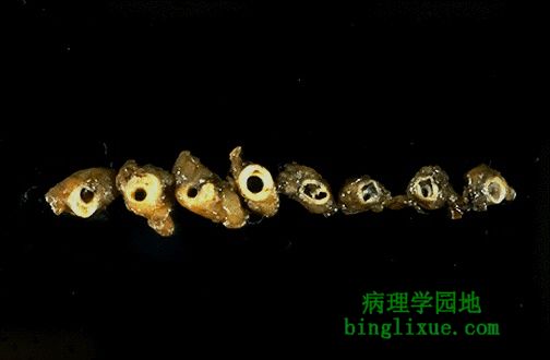 |
These cross sections of the left anterior descending coronary artery demonstrate more pronounced atherosclerosis with narrowing at the left, which is the proximal portion of this artery. Atherosclerosis is generally worse at the beginning of an artery where turbulence is greater. 从左到右依次排列着粥样硬化的左冠状动脉的前降支从近端到远端的横断面,左边的近端冠状动脉内腔更狭窄,粥样硬化更严重。动脉粥样硬化一般是在动脉的起始部位较严重,因为这里湍流更大。 |
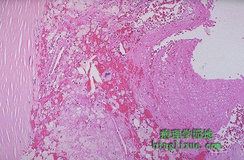 |
This is an atheromatous plaque in a coronary artery that shows endothelial denudation with disruption and overlying thrombus formation at the right. 图示:冠状动脉粥样硬化斑块。内膜断裂剥落,右边伴有血栓形成。
|
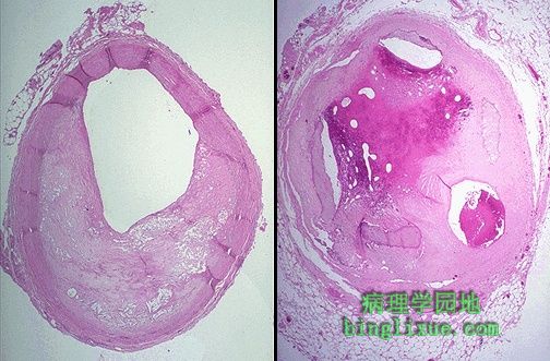 |
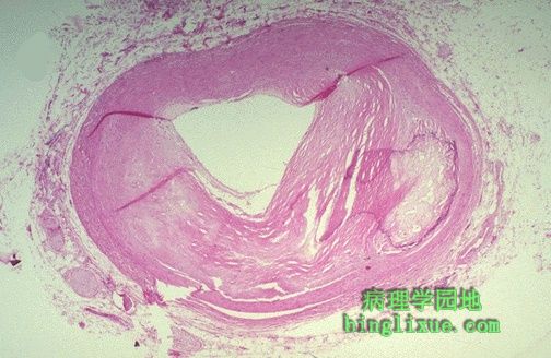
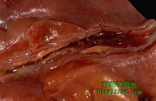
Here is occlusive coronary atherosclerosis. The coronary at the left is narrowed by 60 to 70%. The coronary at the right is even worse with evidence for previous thrombosis with organization of the thrombus and recanalization such that there are three small lumens remaining. 左边发生粥样硬化的冠状动脉内腔狭窄程度达到60%-70%。右边的曾有血栓形成,血栓机化后再通,可以看到有三个内腔。 |
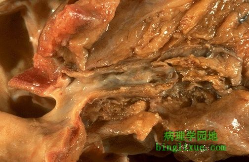 |
This is the gross appearance of severe coronary atherosclerosis, which involves virtually 100% of the surface of the coronary. There is extensive calcification, especially at the right where the lumen is narrowed. 图示:严重的冠状动脉粥样硬化。 冠状动脉表面严重受累,发生了广泛的钙化,尤其是在右边内腔狭窄的部分。 |
 |
Here is a coronary artery with atherosclerotic plaques. There is hemorrhage into the plaque in the middle of this photograph. This is one of the complications of atherosclerosis. Such hemorrhage could acutely narrow the lumen. 图示:冠状动脉粥样硬化斑块。 斑块中有出血,这是动脉粥样硬化的复合病变,可引起急剧的动脉腔狭窄。 |
 |
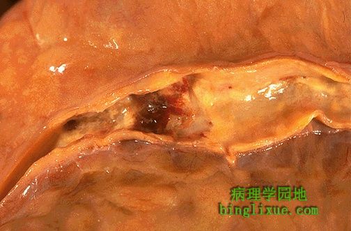
This is coronary thrombosis, one of the complications of atherosclerosis. The dark red thrombus is seen in the anterior descending coronary artery. 图示:冠状动脉粥样硬化形成血栓,是动脉粥样硬化的另一个复合病变。可见冠状动脉前降支有暗红色的血栓形成。 |
 |
Here is the coronary thrombosis at higher magnification. The thrombus occludes the lumen and produces ischemia and/or infarction of the myocardium. 图示:放大后的冠状动脉血栓形成。 血栓使冠脉腔闭塞,引起缺血和(或)心肌梗死。 |