
 |
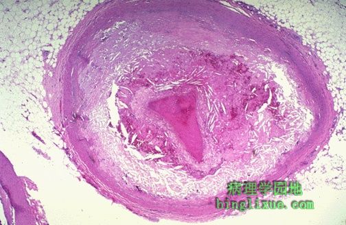
A coronary thrombosis is seen microscopically occluding the remaining small lumen of this coronary artery. 图示:冠状动脉血栓形成使内腔闭塞。 血栓使仅有的很小的内腔闭塞。 |
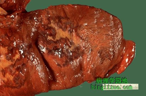 |
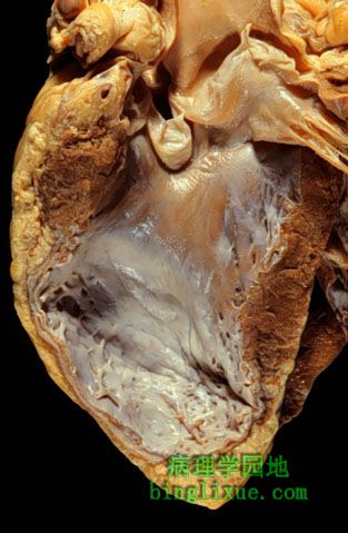
The interventricular septum of the heart has been sectioned to reveal an extensive acute myocardial infarction. The dead muscle is tan-yellow with a surrounding hyperemic border. 心脏室间隔的断面显示了广泛的急性心肌梗死。坏死心肌呈棕黄*色,与周围充血心肌分界清楚。 |
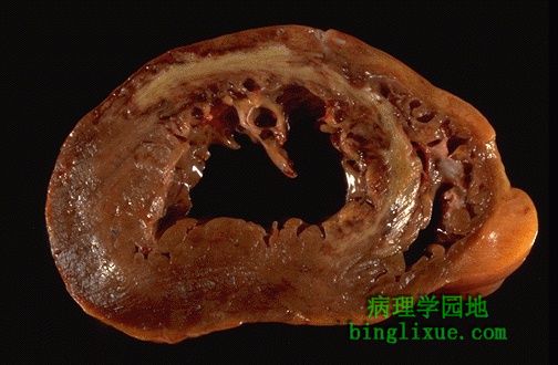 |
This cross section reveals a large myocardial infarction involving the anterior left ventricular wall and septum. 广泛的心肌梗死的断面,发生于左心室壁前面和室间隔。 |
 |
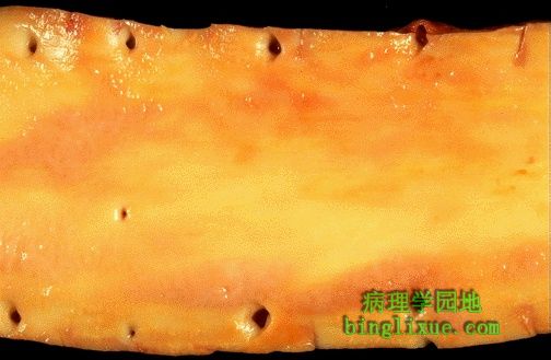
The tan to white areas of myocardial scarring seen from the endocardial surface here represent a remote healed myocardial infarction. 棕褐色到白色的心脏内膜面的心肌显示了陈旧性的心肌梗死。 |
 |
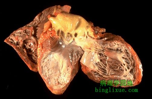
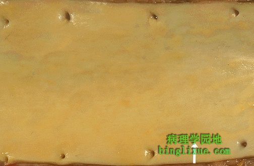
There has been a previous extensive transmural myocardial infarction involving the free wall of the left ventricle. Note that the thickness of the myocardial wall is normal superiorly, but inferiorly is only a thin fibrous wall. The thinned area represents a ventricular aneurysm that has developed as a consequence of the healed infarct. Such an aneurysm represents non-contractile tissue that reduces stroke volume and strains the remaining myocardium. The stasis of blood in the aneurysm predisposes to mural thrombosis. 陈旧性的广泛透壁性左心室心肌梗死。薄的心肌处形成了心室壁瘤。血液淤滞,动脉瘤可引起附壁血栓形成。 |
 |
The pale yellow lipid streaks in the aorta are the earliest lesion of atherosclerosis. 图示:浅黄的脂纹。 它是动脉粥样硬化的早期的病变。 |
 |
Put down that extra slice of pizza and look carefully at this aorta. The white arrow denotes the most prominent fatty streak in the photo, but there are other fatty streaks scattered over the aortic surface. Fatty streaks are the earliest lesions seen with atherosclerosis in arteries. Increased total cholesterol and decreased HDL cholesterol contribute to this process. 图示:动脉粥样硬化的脂质条纹。 可见箭头所指及分散在动脉表面的脂纹。脂纹是动脉粥样硬化的早期病变。总胆固醇的增加和HDL胆固醇的减少可引起该病。 |
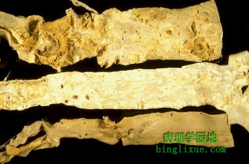 |
Three aortas are shown to demonstrate mild, moderate, and severe atherosclerosis from bottom to top. 从下到上依次是轻度、中度、重度动脉粥样硬化病变。 |
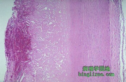 |
Microscopically, the aortic atheromatous plaque is thicker than the remaining media at the right. The plaque contains amorphous pink material with slit-like "cholesterol clefts" of lipid material. There is overlying recent hemorrhage at the left. Thrombus may form on top of such a plaque. 动脉粥样斑块比右边残存的动脉中膜要厚。可见大量针状的胆固醇结晶(针状空隙),左边有新鲜出血,血栓可在这样的斑块顶部形成。 |
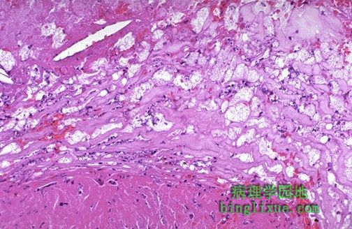 |
At higher magnification, many foam cells (macrophages full of lipid material) and a cholesterol cleft are seen in this atheromatous plaque. 高倍镜下可见粥样斑块中有许多泡沫细胞(即吞噬大量脂质的巨噬细胞)和胆固醇结晶。 |