
 |
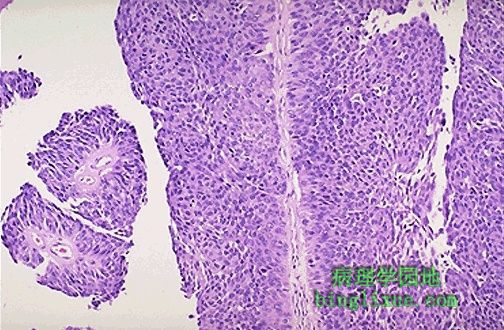
高倍镜下,移行细胞癌的细胞很象尿道上皮,但比正常尿道上皮厚,且呈多形性。 At high power, the urothelial carcinoma does resemble urothelium, but the thickness is much greater than normal and the cells show more pleomorphism. |
 |
肾脏下极的肾细胞癌,相当局限,切面色彩斑驳,有黄*色、白色、棕色区和出血的红色区。尽管肿块通常生长缓慢,但因为腹膜后有相当大的空间,所以在被发现前,肿瘤常常已长到相当大了。对侧的正常肾脏维持着肾脏功能。腰痛、肾区肿块和血尿是三个典型症状。 This is a renal cell carcinoma arising in the lower pole of the kidney. It is fairly circumscribed. The cut surface demonstrates a variegated appearance with yellowish areas, white areas, brown areas, and hemorrhagic red areas. Though these neoplasms are usually slow-growing, they can often reach a considerable size before detection because there is a lot of room to enlarge in the retroperitoneum and there is another kidney to provide renal function. Thus, presenting symptoms and signs usually include flank pain, mass effect, and hematuria. |
 |
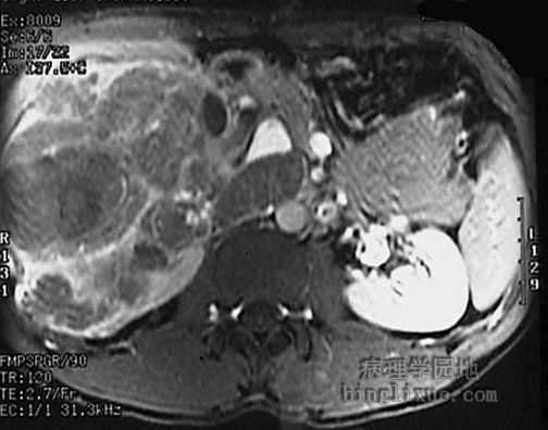
腹部冠状面磁共振(MRI)显示右侧肾下极小的肾细胞癌。 This coronal MRI scan of the abdomen demonstrates a small renal cell carcinoma of the lower pole on the right. |
 |
肾细胞癌非常大,正如15cm标尺所示。一部分正常肾脏突出于正下方。患者本人是一位内科医生。并没有任何早期症状。 This renal cell carcinoma is very large, as indicated by the 15 cm ruler. A portion of normal kidney protrudes at the lower center. This patient was a physician himself and just didn't have any early symptoms. |
 |
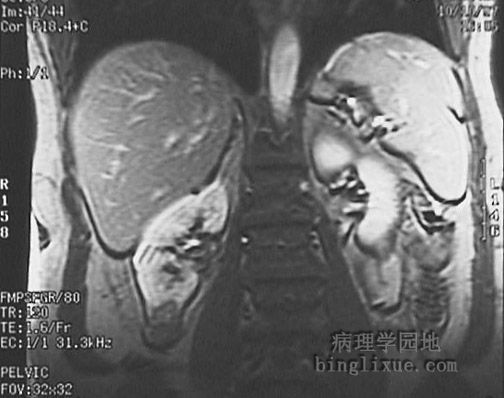
腹部横断面MRI显示右侧大的肾细胞癌。 This transverse MRI scan of the abdomen demonstrates a large renal cell carcinoma on the right. |
 |
肾细胞癌,切面是广泛出血造成的囊肿。 Here is a renal cell carcinoma that on sectioning is mainly cystic with extensive hemorrhage. |
 |
肾细胞癌有侵入肾静脉的倾向,在这个已切除的包有脂肪组织的肾脏,白色的箭头指示肿瘤可侵入腔静脉内,甚至到心脏。但仍可以除去它们。肿瘤蔓延至腔静脉,压迫肾上腺静脉,导致了标本上方的肾上腺出血性梗死。肾细胞癌可以浸润穿过肾包膜。肾细胞癌可转移到其它部位。有近1/4的肾细胞癌首先发现的是转移部位的病变。 Renal cell carcinomas have a tendency to invade into the renal vein, as shown here at the white arrow in a resected kidney surrounded by adipose tissue. They may even crawl up the vena cava and into the heart, but even these can be removed! Here, the tumor extended up the vena cava and occluded the adrenal vein, leading to hemorrhagic adrenal infarction in the adrenal at the top of the specimen. Renal cell carcinomas may invade through the renal capsule. Renal cell carcinomas may metastasize to odd locations, and about a fourth of them first present as metastatic lesions. |
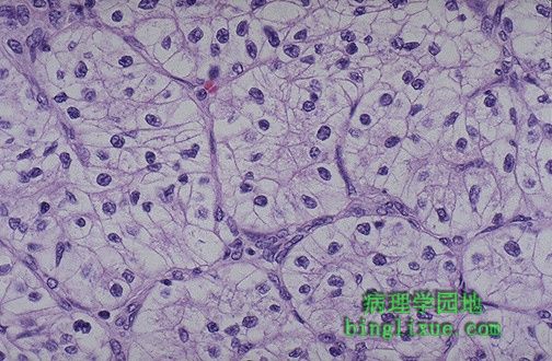 |
典型的肾细胞癌的组织学表现,肿瘤细胞胞浆透亮,呈巢状排列,间质富有毛细血管和血窦,因此称为透明细胞癌。 This is the classic histologic appearance of a renal cell carcinoma: the neoplastic cells have clear cytoplasm and are arranged in nests with intervening blood vessels. This appearance is why they are often called "clear cell carcinomas". |
 |
转移性肾脏肿瘤。是双侧多发性不规则的肿块(有许多因中央坏死而呈中心凹陷或脐样,称癌脐)。肾脏不是肿瘤转移的常见部位。 The multiple irregular bilateral masses (many of which show central indentations, or "umbilications", from necrosis) here represent metastases of carcinoma to the kidneys. Kidney is not a usual site for metastases. |
 |
4岁儿童肾脏,形态较小,含有呈分叶状的灰白色肿块,称为wilms瘤。现在知道,许多wilms瘤的发病和11号染色体的基因缺陷有关。有wilm瘤的儿童常因肿块生长而表现为腹部增大。现在,此病治疗后的5年生存率>90%。 This small kidney from a 4 year old child contains a lobulated tan-white mass. This is Wilm's tumor of the kidney. Many are now known to be associated with genetic defects on chromosome 11. The children with Wilm's tumor usually present with abdominal enlargement from the mass effect. Nowadays, treatment gives a better than 90% 5 year survival. |