
 |
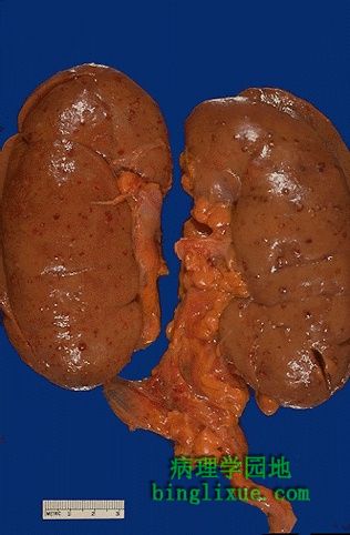
双侧肾脏表面见大量微小脓肿,它们是细菌感染后血行播散造成的。微小脓肿中心是黄*色,外周明显充血。 Here is a kidney with much more The surfaces of both kidneys demonstrate multiple microabscesses from hematogenous spread of a bacterial infection. The microabscesses have yellow centers and prominent hyperemic borders. |
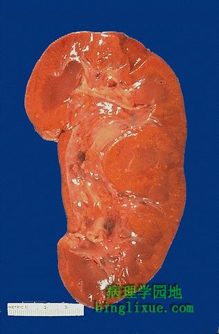 |
肾脏切面上有许多黄*色微小脓肿。 The cut surface of this kidney demonstrates many small yellowish microabscesses. |
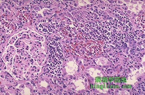 |
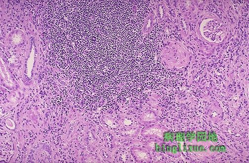
上行性感染导致的急性肾盂肾炎。图中部和右侧的肾小管中充满大量中性粒细胞。 This is an ascending bacterial infection leading to acute pyelonephritis. Numerous PMN's are seen filling renal tubules across the center and right of this picture. |
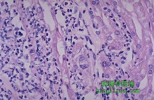 |
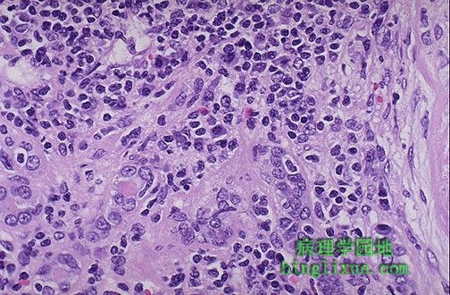
高倍镜下,见急性肾盂肾炎的肾小管和肾间质中有中性粒细胞。 At high magnification, many neutrophils are seen in the tubules and interstitium in a case of acute pyelonephritis. |
 |
一例反复发作的慢性尿路感染病人,见大量慢性炎症细胞。这是慢性肾盂肾炎。 The large collection of chronic inflammatory cells here is in a patient with a history of multiple recurrent urinary tract infections. This is chronic pyelonephritis. |
 |
慢性肾盂肾炎病人高倍镜下见淋巴细胞和浆细胞。在任何慢性肾脏疾病,如肾小球肾炎、肾硬化或肾盂肾炎等,淋巴细胞都很常见。但浆细胞却是慢性肾盂肾炎的特点。 Both lymphocytes and plasma cells are seen at high magnification in this case of chronic pyelonephritis. It is not uncommon to see lymphocytes accompany just about any chronic renal disease: glomerulonephritis, nephrosclerosis, pyelonephritis. However, the plasma cells are most characteristic for chronic pyelonephritis. |
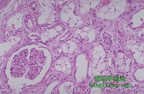 |
乙二醇中毒引起肾小管扩张和空泡状改变。急性肾小管坏死有很多原因,乙二醇中毒是其中的一种。由毒素引起的肾小管坏死通常弥散性累及肾小管,而由缺血(如心衰引起的严重低血压)引起的肾小管坏死则成片状受累。 The tubular vacuolization and dilation here is a result of ethylene glycol poisoning. This is representative of acute tubular necrosis (ATN), which has many causes. ATN resulting from toxins usually has diffuse tubular involvement, whereas ATN resulting from ischemia (as in profound hypotension from cardiac failure) has patchy tubular involvement. |
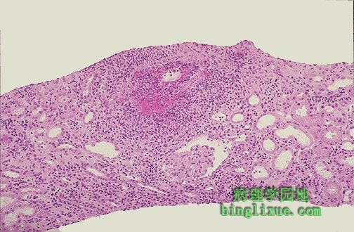 |
肾活检,血管周围有局灶性病变,即血管炎。在正下方是一个正常的肾小球。合格的肾活检最少要有6个肾小球。这样,漏诊局灶性病变的机会较小。 This is a renal biopsy in which there is a focal lesion centered around a blood vessel. Thus, a vasculitis is present. The one glomerulus at the lower center appears normal. An adequate renal biopsy should contain at least 6 glomeruli so that there is less chance that focal lesions will be missed. |
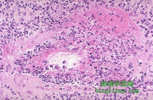 |
高倍镜下可见肾动脉的一个分支有血管炎。这是坏死性肉芽肿性血管炎。本例,血清C-ANCA阳性,诊断为wegener肉芽肿。 At high power, the vasculitis is seen to involve a renal artery branch. This is a necrotizing granulomatous vasculitis. In this case, the C-ANCA serology was positive and a diagnosis of Wegener's granulomatosis was made. |
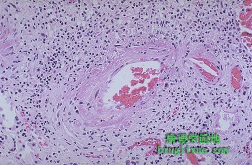 |
肾动脉分支血管炎,淋巴细胞散在分布于血管壁及血管周围。结节性多动脉炎是常累及肾脏的一种系统性血管炎。血清p-ANCA通常是阳性。 Here is a vasculitis of a renal arterial branch. Lymphocytes are scattered in and around the vessel. This happens to be polyarteritis nodosa, a systemic vasculitis that most often affects the kidneys. The P-ANCA serology is usually positive |