
 |
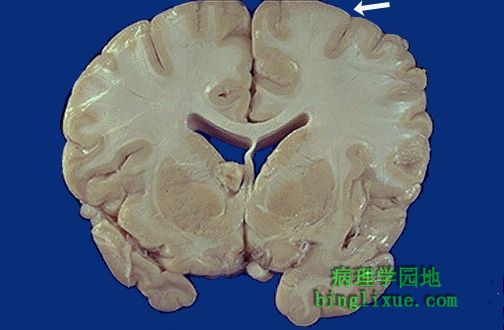
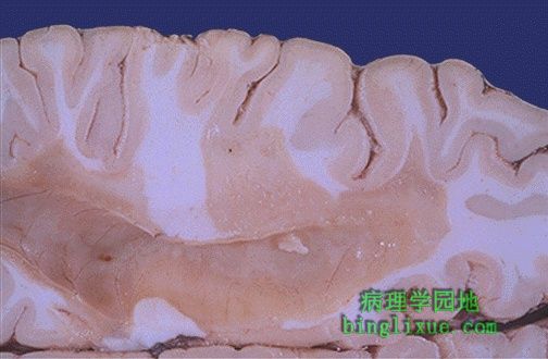
结节性硬化症,常染色体显性遗传,表现为智力低下、癫痫发作等。组织学上的特征表现为出现结节,它是扩大、变硬、变白的脑回。白色的箭头所指即是“结节”。 This is tuberous sclerosis, an autosomal dominant condition characterized by mental retardation and seizures beginning early in life. The characteristic feature is the presence of "tubers" which are enlarged and firm, whitened gyri. A "tuber" is seen at the white arrow. |
 |
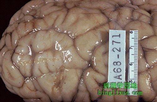
另例结节性硬化症结节。灰质和白质的区别不清。这种病人可能同时患有心脏横纹肌瘤,肾血管肌脂肪瘤,皮肤脂肪腺瘤,胰腺囊肿。 Here is another tuber with tuberous sclerosis. The distinction between grey and white matter is lost. These patients can also have cardiac rhabdomyomas, renal angiomyolipomas, adenoma sebaceum of the skin, and pancreatic cysts. |
 |
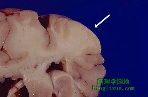
结节性硬化症,变硬变白的脑回比周围正常的脑回宽是其典型表现。 This area of firm, whitened gyri that are broader than surrounding normal gyri is typical for tuberous sclerosis. |
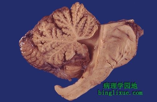 |
慢性酒精中毒病人小脑蚓部前部萎缩。 Here is anterior vermian atrophy of the cerebellum in a patient with chronic alcoholism. |
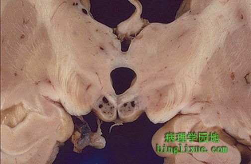 |
乳头体小瘀点性出血为Wernicke病的特征表现,发生于慢性酒精中毒引起的维生素B1缺乏。 The small petechial hemorrhages in the mammillary bodies seen here are characteristic for Wernicke's disease, another complication of chronic alcoholism with thiamine deficiency. |
 |
在白质上看到的是脱髓鞘的大“斑块”,斑块呈灰褐色外观,是多发性硬化症( MS ) 的典型表现。这些斑块导致神经系统功能短暂或逐步丧失的临床症状。因为这种疾病是多病灶的,并且损害随时间改变加重的,所以临床表现也是多种多样。 Seen here in white matter is a large "plaque" of demyelination. The plaque has a grey-tan appearance. Such plaques are typical for multiple sclerosis (MS). These plaques lead to the clinical appearance of transient or progressive loss of neurological function. Since the disease is multifocal and the lesions appear over time, the clinical findings can be quite varied. |
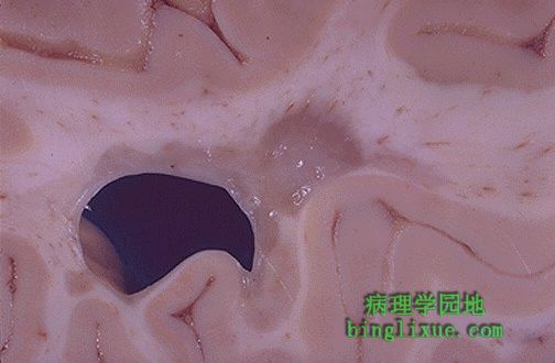 |
脱髓鞘斑块,病人患有多发性硬化症( MS )。采用磁共振(MRI)扫描可发现损害,脑脊液检测结果为免疫球蛋白 G升高,这表明电泳显示的寡克隆带与诊断是非常一致的。 Here is a demyelinated plaque in a patient with multiple sclerosis (MS). The lesions can be seen with MRI scans, but the appearance in the CSF of increased protein from IgG that demonstrates oligoclonal bands on electrophoresis is very consistent with this diagnosis. |
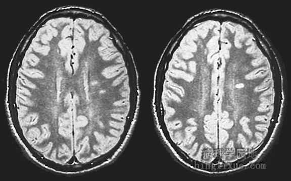 |
两个水平断面的MRI显示在左侧大脑半球的白质区域有三个多发性硬化症斑块。 This FLAIR magnetic resonance imaging (MRI) scan in transverse view at two levels demonstrates three multiple sclerosis plaques in white matter of the cerebral hemisphere on the left. |
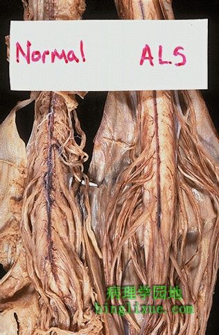 |
肌萎缩侧索硬化( ALS )很少见。它在中年开始发病,并且一直延续数年到死亡。存在前角细胞丧失,因此病人表现为渐进性衰弱, 这可能因神经源性肌萎缩而出现瘫痪。如图示,与正常脊索相比,由于前角细胞丧失,前(腹侧) 脊髓运动神经根萎缩。 Amyotrophic lateral sclerosis (ALS) is uncommon. It begins in middle age and proceeds to death in several years. There is loss of anterior horn cells, so that patients present with progressive weakness that proceeds to paralysis from neurogenic muscular atrophy. Because of the loss of anterior horn cells, the anterior (ventral) spinal motor nerve roots demonstrate atrophy, as seen here in comparison with a normal spinal cord. |
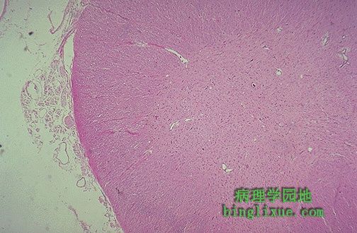 |
脊髓切片上哪里是前角细胞?对于患肌萎缩侧索硬化( ALS )病人来说,不可能看到前角细胞。 Where are the anterior horn cells in this section of spinal cord? They are absent in a patient with amyotrophic lateral sclerosis (ALS). |