
 |
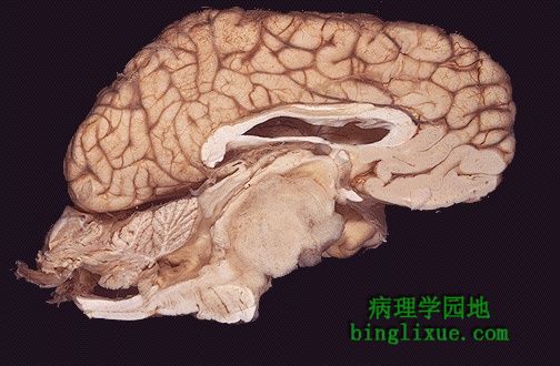
脑干矢状面显示一个较大的脑干胶质瘤 。大多数胶质瘤是星形细胞瘤。 This sagittal section of brain demonstrates a large brainstem glioma. Most gliomas are astrocytomas. |
 |
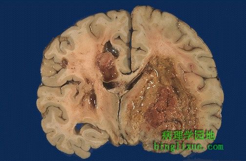
胶质瘤发生于大脑半球。对大多数胶质瘤,要分清其边界很困难。 This glioma is arising in the cerebral hemisphere. As in most gliomas, it is difficult to tell where the margin is. |
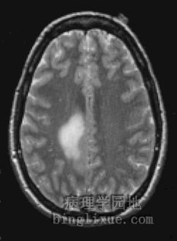 |
T2加权横断面MRI显示浸润性肿块累及右额叶中后部和顶叶,病变与胶质瘤相一致。 This T2 weighted magnetic resonance imaging (MRI) scan in transverse view demonstrates an infiltrative mass involving the posteromedial right frontal lobe and parietal lobe, consistent with a glioma. |
 |
低倍镜,与右边临近的脑组织相比,左边神经胶质瘤细胞体积大并有多形性,边界不清。 At low power, a glioma at the left shows greater cellularity and pleomorphism than adjacent brain at the right, but the margin is not distinct. |
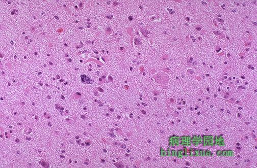 |
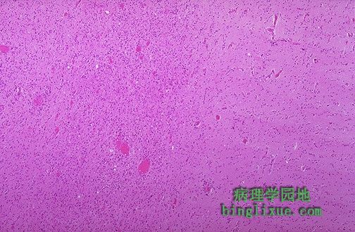
与正常大脑相比,星形细胞瘤显示出细胞内容丰富并有明显多形性。注意中心部位的多形性细胞。 This astrocytoma demonstrates increased cellularity and pleomorphism, as compared to normal brain. Note the very pleomorphic cell in the center. |
 |
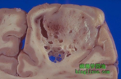
胶质瘤中最糟的类型--多形性胶质母细胞瘤( GBM )。肿瘤在坏死和出血区域血管相当丰富。注意肿瘤如何穿过正中线到对侧的大脑半球。 This is the worst possible form of glioma--a glioblastoma multiforme (GBM). These neoplasms are quite vascular with prominent areas of necrosis and hemorrhage. Note how this one has crossed the midline to the opposite hemisphere. |
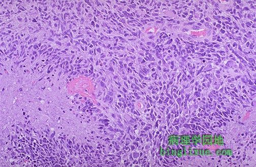 |
多形性胶质母细胞瘤( GBM )显示肿瘤细胞深染和多形性。注意典型的血管构成,如图左边坏死区域肿瘤细胞呈栅栏状围绕。 This glioblastoma multiforme (GBM) demonstrates marked cellularity with marked hyperchromatism and pleomorphism. Note the prominent vascularity as well as the area of necrosis at the left with neoplastic cells palisading around it. |
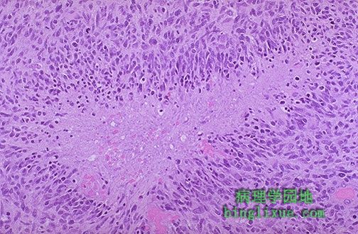 |
多形性胶质母细胞瘤( GBM )肿瘤细胞假栅栏状坏死,肿瘤细胞广泛浸润,特别是沿着白质,甚至穿过脑脊液。 Here is another example of pseudopalisading necrosis of neoplastic cells in a glioblastoma multiforme (GBM). The cells of a GBM can infiltrate widely, particularly along white matter tracts, and even through the CSF. |
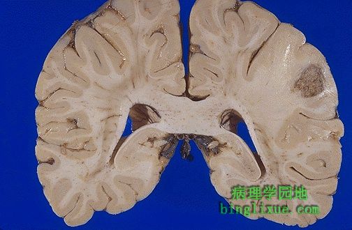 |
左边(图右侧)为肺癌的脑转移。转移大多数出现在大脑中动脉分布区的灰质和白质的分界处,如图所示。 At the left is seen a metastasis from a lung carcinoma. Metastases most often appear at the border of the grey and white matter in the distribution of the middle cerebral artery, as in this case, because that is where the blood flow (vascular distribution) is most likely to take metastases. |