
 |
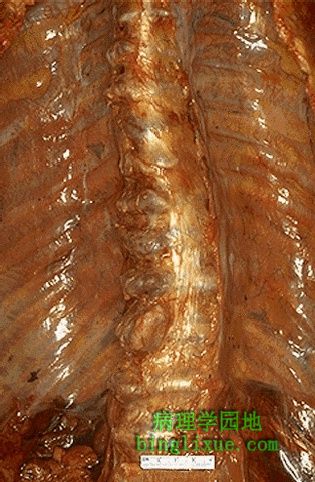
脊椎显著“唇形变”的退行性骨关节炎。骨关节炎在50岁以上人群中随年龄而增加。主要累及大关节,比如髋关节或膝关节。骨关节炎源于关节软骨的磨损破坏甚至丧失。也可见有个向右的弯曲,表明存在轻微脊柱侧凸。 Here is another example of degenerative osteoarthritis in which there is prominent "lipping" of the vertebrae. Osteoarthritis increases with age beyond 50. Generally, a few large joints are involved, such as hip or knee. Osteoarthritis is due to wear and tear with loss of articular cartilage. Note also that there is a curve to the right indicating a minor degree of scoliosis. |
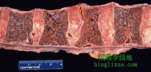 |
椎骨明显的骨质疏松症,可见骨小梁薄弱甚至丧失。从右侧数第二块椎骨显示出更明显的椎体受压。骨质疏松症随年龄增长出现加速性骨质丧失且在绝经后妇女中特别常见,使其有骨折的风险(如髋骨、腕骨、椎骨)。 The bone in these vertebral bodies demonstrates marked osteoporosis with thinning and loss of bony trabeculae. The second body from the right shows a greater degree of compression than the others. Osteoporosis is accelerated bone loss with age and is particularly common amongst postmenopausal women, putting them at risk for fractures (hip, wrist, vertebrae). |
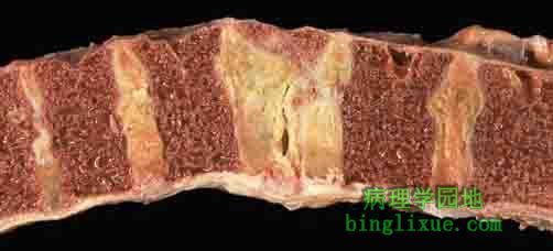 |
脊柱压缩性骨折。中间椎体明显减小。这种骨折常见于骨质疏松,尤其是老年妇女骨质疏松时存在加速性骨组织破坏丧失。骨折甚至在微小创伤作用下就可发生。 Here is a "compressed" fracture of the vertebral column. The middle vertebral body shown here is greatly reduced in size. Such fractures are common in persons with osteoporosis in which there is accelerated bone loss, particularly older women, and can occur with even minor trauma. |
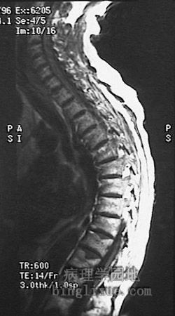 |
脊柱MRI显示明显的脊柱后凸,也可见伴有压缩性骨折。主要是由于长期骨质疏松形成的。 This MRI of the spine demonstrates marked kyphosis with compressed fractures. Such a finding can be seen as a consequence of osteoporosis. |
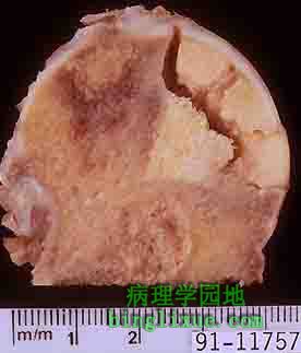 |
可见股骨头右上方楔形的缺血性坏死(骨坏死)区。缺血性坏死源于骨缺血,而引起骨缺血的原因有多种,包括创伤与皮质类固醇给药,可是原因不明的情况也普遍。可由活动时疼痛渐渐发展为休息时疼痛。最后,坏死骨塌陷,扭曲叠压关节软骨从而产生继发性骨关节炎。 Note the wedge-shaped area of avascular necrosis (osteonecrosis) at the upper right of this femoral head. Avascular necrosis results from bone ischemia, which can be due to many causes, including trauma and corticosteroid administration, though idiopathic cases are common. There is pain with activity, progressing to pain at rest. Eventually, the necrotic bone collapses, distorting the overlying articular cartilage and producing secondary osteoarthritis. |
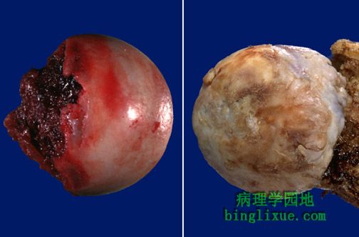 |
左边的股骨头(骨折离断)显示了光滑、闪亮的关节软骨,而右边的股骨头则显示了骨关节炎的典型的粗糙、坚硬、不规则的外形。 The femoral head at the left (removed because of fracture) shows smooth, glistening articular cartilage, while the femoral head at the right shows a rough, eburnated, irregular appearance typical for osteoarthritis. |
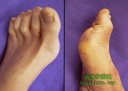 |
图示足畸形为拇囊炎,表现为MP关节隆起。畸形使行走更困难,且要是拇囊炎使鞋不合适还可能产生厚胼胝体或溃疡形成。这种损害可通过外科矫治减弱。 The deformity of the foot shown here is known as a "bunion" and represents protrusion of the MP joint. Such a deformity makes walking more difficult and can produce a thick callus or ulceration when the bunion makes shoes ill-fitting. This lesion can be lessened by surgical correction. |
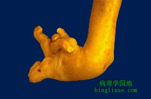 |
类风湿关节炎(RA)引起的手畸形。这种自身免疫病导致滑膜增生与关节破坏,典型病变为对称性畸形,出现于手与足的小关节,其次是腕关节、踝关节、肘关节、膝关节。血清学检测,大部分(并非全部)病人可查到类风湿因子。 This deformity of the hand is due to rheumatoid arthritis (RA). This autoimmune disease leads to synovial proliferation and joint destruction, typically in a symmetrical pattern involving small joints of hands and feet, followed by wrists, ankles, elbows, and knees. Rheumatoid factor can be identified serologically in most, but not all, RA patients. |
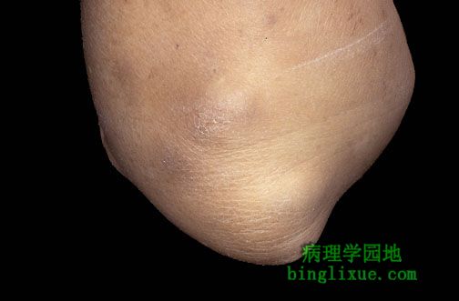 |
有时类风湿关节炎(RA)病人在皮下压痛点处有类风湿结节形成,如图示的肘。类风湿结切也可见于内脏,如肺胸膜上。 Sometimes persons with rheumatoid arthritis (RA) have rheumatoid nodules form in subcutaneous locations at pressure points, such as the elbow shown here. Rheumatoid nodules may also appear viscerally, such as on the pleura of the lung. |
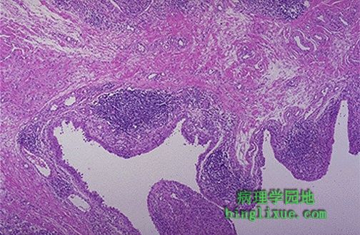 |
类风湿关节炎滑膜。淋巴细胞与浆细胞浸润的慢性炎症使增生性结节下方呈现为蓝色区域。滑膜增厚,形成许多绒毛样突起,即血管翳,血管翳具有破坏性,可侵蚀关节软骨,最终破坏关节。 This is the synovium in rheumatoid arthritis. There is chronic inflammation with lymphocytes and plasma cells that produce the blue areas beneath the nodular proliferations. This "pannus" is destructive and produces erosion of the articular cartilage, eventually destroying the joint. |