
 |
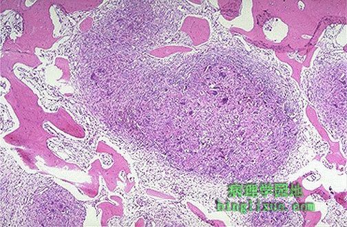
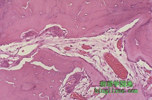
甲状旁腺功能亢进病人骨棕色瘤。高甲状旁腺激素水平提高了破骨细胞的活性并导致不规则的骨溶解吸收,伴随微骨折、出血、巨噬细胞增殖及纤维性结缔组织增生。 Here is a "brown tumor" of bone in a patient with hyperpara-thyroidism. The high parathormone levels increase osteoclast activity and produce irregular bone resorbtion with microfractures and hemorrhage and macrophage proliferation and fibrous connective tissue proliferation. |
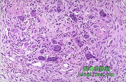 |
棕色瘤中心包含破骨细胞、单核细胞、成纤维细胞,并伴有局灶性出血。出血形成的血铁黄素使病变肉眼观察呈深棕色。现今这种病变不常见,因为它出现之前甲状旁腺功能亢进已被治愈。 The center of the "brown tumor" contains osteoclasts and mononuclear cells and fibroblasts with focal hemorrhages. The hemosiderin from the hemorrhage produces the grossly brown color. Such lesions are nowadays uncommon because hyperparathyroidism is treated before such lesions develop. |
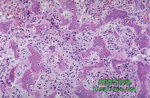 |
图示由不规则反应性新生骨组成的骨样骨瘤。骨样骨瘤常见于十几岁男性中轴骨骼(尤其是胫骨和股骨)的骨皮质。成骨细胞瘤恰是椎骨上的大骨样骨瘤。为良性肿瘤,可通过切除治愈。 This is the central nidus of an osteoid osteoma composed of irregular reactive new bone. Osteoid osteomas usually occur in the axial skeleton (especially tibia and femur) in bone cortex of young males in the second decade of life. An osteoblastoma is just a big osteoid osteoma in the vertebra. These lesions are benign and cured by resection. |
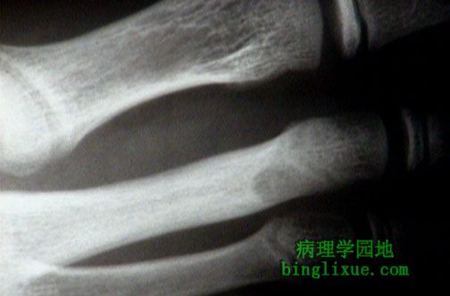 |
骨样骨瘤,X线可见跖骨骨皮质的小圆中央透亮区被硬化骨所包绕。 This is the central nidus of an osteoid osteoma. Radiographically, there is a small round central lucent area in the metatarsal bone cortex surrounded by sclerotic bone. |
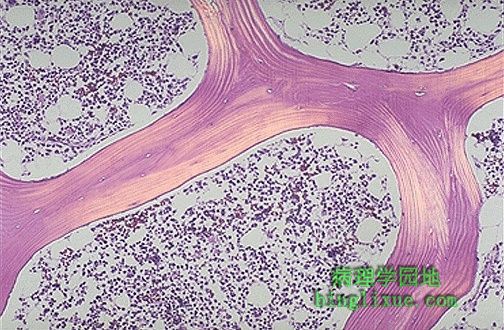 |
图示偏振光显微镜下的正常骨松质,可见突出的板层状结构。骨板连续均一,偶尔可见包含骨细胞的腔隙。可见蜂窝状骨髓位于骨板之间。 Here is normal cancellous bone as seen under polarized light microscopy, which highlights the lamellar structure. The bony spicules are even, with occasional lacunae containing osteocytes. Cellular marrow is seen between the spicules of bone. |
 |
Paget病病人混合破骨-成骨期的骨。一行成骨细胞正在中右部形成新骨,而腔隙内的多核破骨细胞正在中左与中底部破坏骨。结果形成了不均一的板层结构的骨的不协调嵌合体。此期的前一期为溶解期,紧接着为硬化期。 This is Paget's disease of bone in which the mixed osteoclastic-osteoblastic stage is present. A line of osteoblasts is present a the center-right forming new bone, but lacunae containing multinucleate osteoclasts are at the center left and lower center destroying bone. The result is a patchwork mosaic of bone without an even lamellar structure. This stage is preceded by a mainly lytic phase and is followed by a "burnt out" sclerotic phase. |
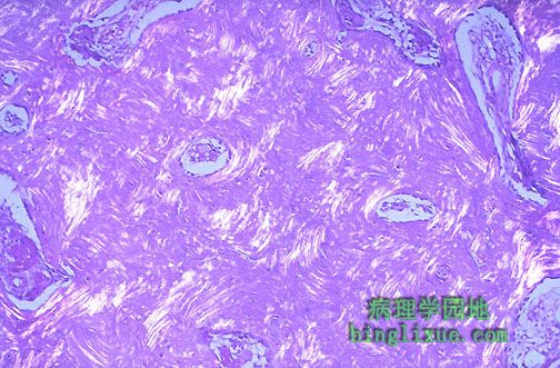 |
paget病见于老年欧洲高加索人种。在偏振光下,不规则骨板清楚可见。 Paget's disease is seen in elderly Caucasians of European ancestry. Under polarized light, the irregularities of the bony lamellae are apparent. |
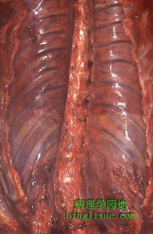 |
图示脊柱弯曲为脊柱侧凸。本病例中下腰部向左侧弯曲不明显,病人看上去不会得并发症。可能发展为更明显的侧弯并导致严重畸形。 The curvature of the vertebral column seen here is known as scoliosis. In this case, the curve to the left in the lower lumbar region is not pronounced, and this person will be unlikely to have complications. Larger curves can progress and cause serious deformity. |
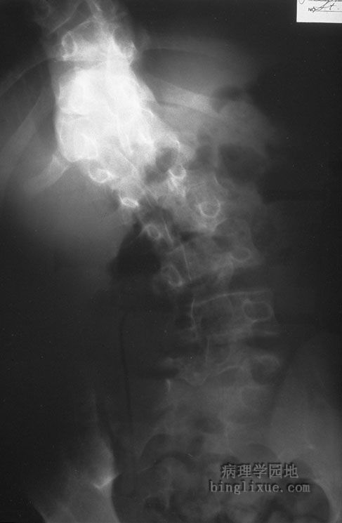 |
X线显示典型的脊柱侧凸。 Curvature of the vertebral column is demonstrated here, typical for scoliosis. |
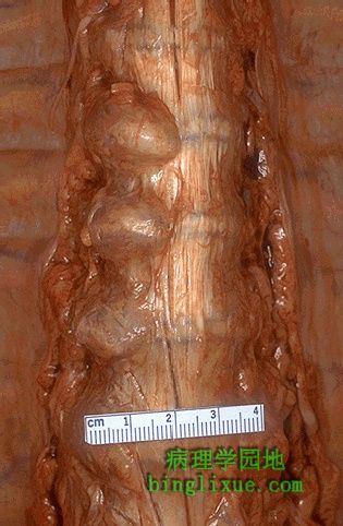 |
左边脊柱多节的赘生物是由退变性骨关节炎引起的。这是椎体明显的“唇形变”。 The knobby excrescences at the left side of this vertebral column are due to degenerative osteoarthritis. This is the pronounced "lipping" of the vertebral bodies. |