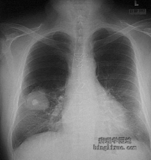
胸部X线显示右肺下叶直径达5cm的鳞状细胞癌,其中直径1.5cm的高密度区是肿瘤发生后的钙化的肉芽肿。胸骨见到的钢丝圈是原来冠脉搭桥术时留下的。
This chest radiograph demonstrates a large 5 cm diameter squamous cell carcinoma of the right lower lobe. The 1.5 cm bright opacity in the middle of the mass is a calcified granuloma that was seen on lateral view to be behind the neoplasm. Additional calcified granulomatous areas are medial to the mass. The sternal wire loops are from a previous coronary artery bypass procedure.
