
 |
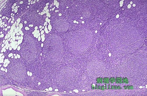
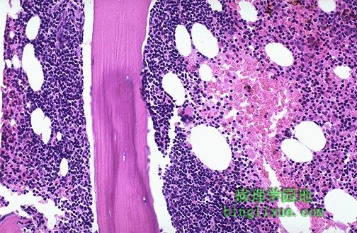
淋巴瘤病人淋巴结,以淋巴样肿瘤细胞增生形成瘤为典型的恶性过程。淋巴结被膜被侵袭,淋巴瘤的细胞已经侵入周围的脂肪组织。淋巴滤泡增多、大小不一。属恶性淋巴瘤,小裂细胞型,滤泡性(也称为:恶性淋巴瘤,低分化淋巴细胞型,结节性)。 Here is a lymph node involved by lymphoma, a malignant process characterized by the proliferation of neoplastic lymphoid cells. The capsule of the node has been invaded and the lymphomatous cells extend into the surrounding adipose tissue. Note that the follicles are numerous and irregularly sized. This is a malignant lymphoma, small cleaved cell type, follicular (also known as: malignant lymphoma, poorly differentiated lymphocytic type, nodular). |
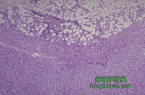 |
恶性淋巴瘤呈弥散性,不见淋巴滤泡。低倍镜下已经看不到正常的淋巴结结构。淋巴结被小的淋巴瘤细胞浸润,浸润已经延伸到淋巴结的囊,浸润到周围的脂肪组织。诊断是:恶性淋巴瘤,小淋巴细胞型,弥漫性(也称为:“高分化”淋巴细胞淋巴瘤。 This pattern of malignant lymphoma is diffuse and no lymphoid follicles are identified. Under low power, note that the normal architecture of the lymph node is obliterated. The lymph node is replaced by an infiltrate of small (mature-appearing) neoplastic lymphocytes, and the infiltrate extends through the capsule of the lymph node and into the surrounding fat. The diagnosis is: malignant lymphoma, small lymphocytic type, diffuse (also known as: "well-differentiated" lymphocytic lymphoma). |
 |
染色体组型为46,XY,t(8;14)显示Burkitt 型(小无裂细胞型)淋巴瘤典型的异位。此型淋巴瘤非洲多见,常见于儿童。在美国,儿童和年轻人很少见,主要累及腹部。细胞生成分析在区分很多恶性血液病方面起重要作用。 This karyotype of 46, XY, t(8;14) demonstrates the translocation typical for a Burkitt type (small non-cleaved) lymphoma. This is one of the most common lymphomas in Africa and most often appears in children. In the U.S., it is less commonly seen in children and young adults and involves the abdomen. Cytogenetic analysis is useful in characterizing many hematologic malignancies. |
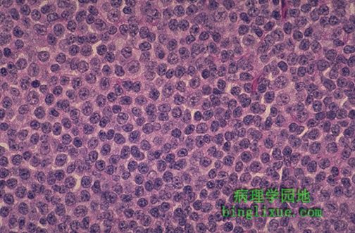 |
许多发生于成年人的非霍金氏淋巴瘤是大细胞淋巴瘤,如图中倍镜示。可能与免疫抑制状态有关(例如AIDS),是典型B细胞起源的肿瘤。细胞体积大,有明显的核和丰富的细胞质。早期趋向于局部化,但随后迅速扩大,比较低级的淋巴瘤有更大的侵袭淋巴结外组织的倾向。 Many non-Hodgkin's lymphomas seen in adults are large cell lymphomas such as the one here at medium power, but they can be associated with immunosuppressed states (such as AIDS), and are typically of B cell origin. The cells are large, with prominent nucleoli and abundant cytoplasm. This disease tends to be localized (low stage), but with more rapid enlargement, and a greater propensity to be extranodal than the low grade lymphomas. |
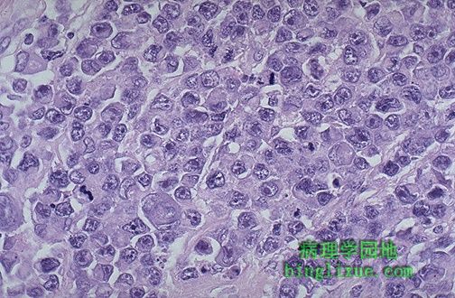 |
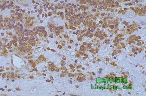
此恶性淋巴细胞体积大、有较丰富的细胞浆,核圆型或卵圆型,核仁明显,偶有核分裂像,诊断为弥漫大B细胞淋巴瘤(也称免疫母细胞淋巴瘤)。主要需要鉴别的是转移癌。免疫过氧化物酶技术检测,若存在单克隆免疫球蛋白将有助于确定恶性淋巴瘤。抗原CD19和CD20有助于确定为B细胞起源。 The malignant lymphocytes here are very large with a moderately abundant cytoplasm, and the nuclei are round to ovoid with prominent nucleoli and occasional mitoses. The diagnosis is diffuse large B cell lymphoma (also known as immunoblastic lymphoma). The major differential diagnosis in this case would be a metastatic carcinoma. The presence of monoclonal immunoglobulin as demonstrated by immunoperoxidase technique would help to confirm this lesion as a malignant lymphoma. Demonstration of CD19 and 20 antigens would classify it as B cell in origin. |
 |
骨髓活检可以揭示恶性淋巴瘤。图示骨小梁周围小兰细胞(淋巴瘤细胞)浸润。 A bone marrow biopsy can reveal malignant lymphoma. Here, there is a peritrabecular infiltrate of small blue cells which is the lymphomatous infiltrate. |
 |
器官移植尤其是心脏移植(当然也包括少量肾移植和骨髓移植)后,免疫抑制剂治疗可促进EB病毒感染T细胞。图示免疫过氧化物酶染色可见EB病毒,是移植后淋巴组织增生症(PTLD),它象淋巴瘤,但免疫抑制减弱时可消退。 Following organ transplantation, particularly for heart, but also to a lesser extent with kidney and bone marrow, immunosuppressive therapy may promote an expansion of Epstein-Barr virus (EBV) infected T-cells, seen here with immunoperoxidase staining for EBV. This is a post-transplantation lymphoproliferative disorder (PTLD), which acts like a lymphoma, but will recede when immunosuppression is diminished, if possible. |
 |
颅骨显示多发性骨髓瘤典型的圆形“打孔样”病损。 The skull demonstrates the characteristic rounded "punched out" lesions of multiple myeloma. |
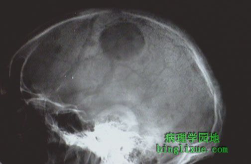 |
多发性骨髓瘤的圆形“打孔样”病损,图示颅骨X线显示为一半透明区域。 The rounded "punched out" lesions of multiple myeloma appear as lucent areas with this skull radiograph. |
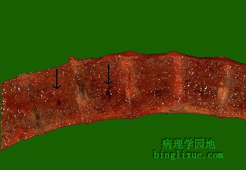 |
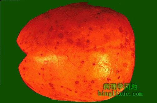
圆形病灶充满柔软的粉红色物质,图示的脊椎骨切面显示骨髓瘤病灶。 Round lesions filled with a soft reddish material are indicative of foci of myeloma in this section of vertebral bone. |