
 |
The liver shows a small abscess here filled with many neutrophils. This abscess is an example of localized liquefactive necrosis. 肝脏小脓肿,充满了嗜中性粒细胞。该脓肿是局部性液化性坏死的典型例子。 |
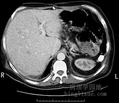 |
This magnetic resonance imaging (MRI) scan of the abdomen in transverse view demonstrates multiple small lucent lesions representing abscesses from septicemia. The spleen is absent. MRI(磁共振成像)扫描腹部横断面显示的多发性小透亮区为脓毒症病人发生的小脓肿。 |
 |
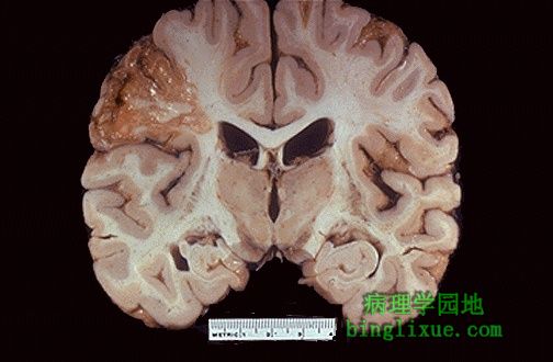
This is liquefactive necrosis in the brain in a patient who suffered a "stroke" with focal loss of blood supply to a portion of cerebrum. This type of infarction is marked by loss of neurons and neuroglial cells and the formation of a clear space at the center left. 图示:脑组织液化性坏死。 该病人是脑血流减少使脑组织一部分发生了缺血性坏死。左侧中央的明亮的坏死区神经元和神经胶质细胞消失。 |
 |
At high magnification, liquefactive necrosis of the brain demonstrates many macrophages at the right which are cleaning up the necrotic cellular debris. The job description of a macrophage includes janitorial services such as this, particularly when there is lipid. 高倍镜下,在右侧的脑组织液化性坏死灶中许多巨噬细胞清除了坏死的细胞碎片。巨噬细胞的这种防御功能,尤其是在含脂质较多的组织中发挥得更明显。 |
 |
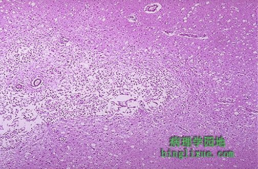
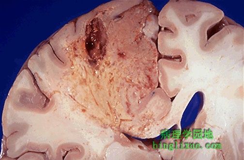
Grossly, the cerebral infarction at the upper left here demonstrates liquefactive necrosis. Eventually, the removal of the dead tissue leaves behind a cavity. 肉眼观:左上角的脑组织梗死表现为液化性坏死。最终坏死组织被清除后留下空洞。 |
 |
As this infarct in the brain is organizing and being resolved, the liquefactive necrosis leads to resolution with cystic spaces. 图示:脑组织梗死液化 脑组织梗死灶正在进行机化并且逐渐被溶解液体吸收后往往形成囊腔。 |
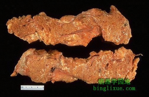 |
This is fat necrosis of the pancreas. Cellular injury to the pancreatic acini leads to release of powerful enzymes which damage fat by the production of soaps, and these appear grossly as the soft, chalky white areas seen here on the cut surfaces. 图示:胰腺脂肪坏死 胰腺腺泡细胞损伤释放溶解酶,产生钙皂破坏脂肪组织,从而使相应部位呈现白色。 |
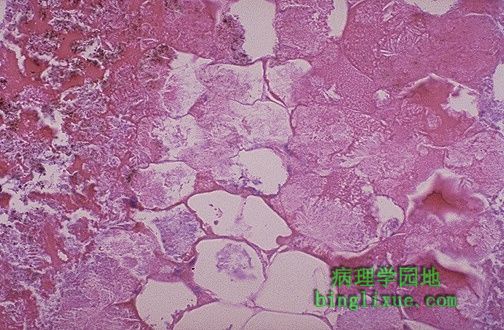 |
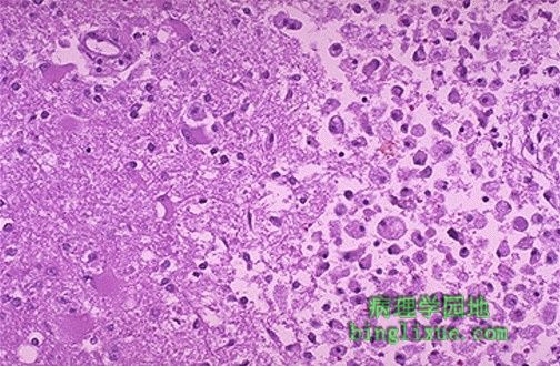
Microscopically, fat necrosis is seen here. Though the cellular outlines vaguely remain, the fat cells have lost their peripheral nuclei and their cytoplasm has become a pink amorphous mass of necrotic material. 镜下:脂肪坏死 虽然脂肪细胞轮廓隐约可见,但细胞边缘的细胞核已消失,细胞浆也变成了结染无结构的坏死物。
|
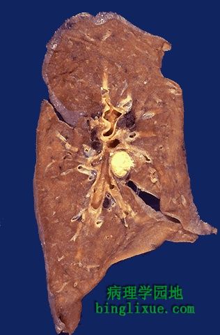 |
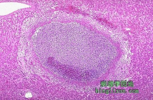
This is the gross appearance of caseous necrosis in a hilar lymp node infected with tuberculosis. The node has a cheesy tan to white appearance. Caseous necrosis is really just a combination of coagulative and liquefactive necrosis that is most characteristic of granulomatous inflammation. 图示:肺结核病肺门淋巴结干酪样坏死 呈黄褐色到白色外观,是凝固性坏死和液化性坏死的复合性坏死,属于炎症肉芽肿的特征性变化。 |
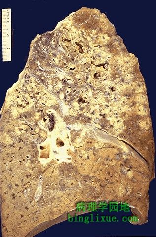 |
This is more extensive caseous necrosis, with confluent cheesy tan granulomas in the upper portion of this lung in a patient with tuberculosis. The tissue destruction is so extensive that there are areas of cavitation (cystic spaces) being formed as the necrotic (mainly liquefied) debris drains out via the bronchi. 图示:肺结核广泛干酪样坏死 肺上部伴有广泛的黄褐色干酪样肉芽肿形成,组织破坏严重,坏死物分解液化后形成许多小空洞,坏死碎片通过支气管排出。 |