
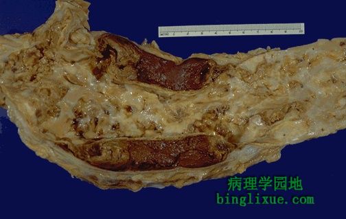
主动脉纵向剖开可以清楚地看到局部结构。剖面图的两端可见红褐色的血栓在主动脉内延伸。左边的内膜已经破裂,使得主动脉形成了一个“双管腔”。见重度动脉粥样硬化合并囊状中层坏死以及高血压,增加了剖开术的危险性。
This aorta has been opened longitudinally to reveal an area of fairly limited dissection that is organizing. The red-brown thrombus can be seen in on both sides of the section as it extends around the aorta. The intimal tear would have been at the left. This creates a "double lumen" to the aorta. This aorta shows severe atherosclerosis which, along with cystic medial necrosis and hypertension, is a risk factor for dissection.